Summary
Hyperpigmentation can be common with full-thickness skin grafts that have prolonged healing. Dermabrasion and/or laser therapy is very efficacious in improving final appearance.
Hypopigmentation is often unpredictable in occurrence and difficult to correct.
The treatment of contour abnormalities begins with effective scar management including massage, silicone sheeting, and intralesional triamcinolone. If contour abnormalities are not resolved with final scar maturation, then revision surgery is indicated.
Although rare in postoperative Mohs patients, practitioners must remain vigilant about the possibility of cancer recurrence.
22.1 General Considerations
The management of complications in the late healing stage involves poor color match of the flap or graft, poor contour of the flap or graft, unexpected hypertrophic or keloid scarring of the flap or graft, as well as functional issues such as nasal airway obstruction or lip ectropion and then finally cancer recurrence after the initial resection. Unfortunately, the management of late complications also encompasses the management of suboptimal results from the patient’s perspective. These can either be true suboptimal surgical results or failure of the patient’s expectations matching expected surgical results. Patient education can obviate and minimize many of the expected results. A perfectly fine and acceptable, even very good, surgical result can be viewed by the patient as an abject disaster if their expectations do not match the surgical reality. It is foolish of the surgeon and of low yield to continue to point out “how good” the results are if the patient remains unhappy. Education with time spent discussing the procedure, as well as the patient’s perception of the results that include representative postoperative photographs, is time well spent. Additionally, a second opinion can also help temper this often difficult clinical situation.
22.1.1 Hypo- and Hyperpigmentation
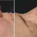
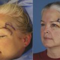
The management of color complications can be hypo- or hyperpigmentation. More commonly, hyperpigmentation with healed full-thickness skin grafts or flaps is often the result of continued surface desquamation despite adequate graft take, and this will require multiple episodes of dermabrasion to improve the final color. 1, 2 Laser has some proven benefit and pulsed-dye laser can commence as early as 6 weeks after graft inset, often in conjunction with dermabrasion. The management of hypopigmentation is more difficult and its occurrence is somewhat unpredictable even with good graft take. Well-vascularized healthy grafts may have a period of transient hypopigmentation that corresponds to the absent vasomotor function, but this will be restored as the vasomotor function returns. Unfortunately, some hypopigmentation will never resolve and long-term management of can be difficult (▶ Fig. 22.1 and ▶ Fig. 22.2).
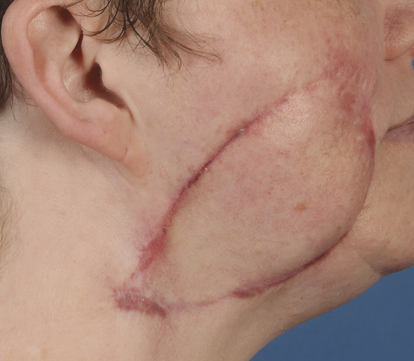
Fig. 22.1 Hyperpigmentation.
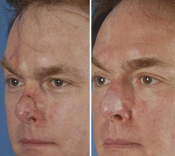
Fig. 22.2 Hypopigmentation.
22.1.2 Contour Abnormalities
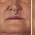
Unacceptable contour abnormalities are initially managed with rigorous postoperative scar therapy including silicone sheeting and massage. 3, 4 If there is a hypertrophic scar component to this, then intralesional triamcinolone on a scheduled injection series is added to the treatment protocol. 5, 6 No attempt is made to revise contour abnormalities until the graft or flap repair has entered the supple healing stage and the skin is pliable. Depressed contour abnormalities in the late healing stage are fairly easily managed by flap re-elevation and placement of a dermal fat graft, cartilage graft, or fat grafting. 7, 8, 9 The management of hypertrophic or keloid scars depends on the accurate identification of either. Keloid scars can be tremendously difficult to manage and with simple excision alone or simple steroid injection alone show an unacceptable (as high as 90%) recurrence rate. 6, 10, 11 Multimodal therapy, including surgical excision and intralesional steroids with or without mitomycin, is the only acceptable management for keloid therapy. 6, 10, 11 Recurrent keloids are best managed by multimodality therapy that includes surgical excision and immediate short-term radiation therapy. 6, 10, 11 Simpler to manage are hypertrophic scars and the management should include a patient discussion that everyone heals along a continuum and hypertrophic scars are a normal portion of this continuum. Aggressive management should include silicone sheeting, scar massage, as well as scheduled intralesional steroid therapy 10, 12 (▶ Fig. 22.3).
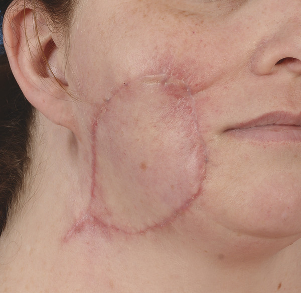
Fig. 22.3 Hypertrophic scars.
Related posts:
Stay updated, free articles. Join our Telegram channel

Full access? Get Clinical Tree


