Sling Procedures for Stress Urinary Incontinence

16 Sling Procedures for Stress Urinary Incontinence


HISTORICAL PERSPECTIVES


The vaginal approach for the surgical management of urodynamic stress urinary incontinence (SUI) associated with urethral hypermobility and intrinsic sphincter deficiency (ISD) has been comprised of a wide variety of procedures based on different surgical principles. Historically, suburethral sling procedures have been reserved for patients with severe stress incontinence, previous surgical failures, and patients who have significant ISD. Until recently, anterior colporrhaphy with Kelly’s plication and transvaginal needle suspension procedures were used for the transvaginal repair of primary stress incontinence and associated urethral hypermobility. Numerous studies, however, have shown that the objective success rate after anterior colporrhaphy and suburethral plication is significantly less than what could be achieved with retropubic colposuspension or a suburethral sling procedure. For this reason, suburethral plication should no longer be considered as a procedure for the correction of stress incontinence and should only be performed as part of an anterior colporrhaphy or cystocele repair without incontinence or in the occasional elderly patient with mild incontinence in whom surgical morbidity should be kept low (see Chapter 19).


Transvaginal needle suspension procedures were first described by Pereyra in 1959. The needle urethropexy underwent more than 20 modifications in an attempt to improve the cure rates and minimize complications. Modifications involved various amounts of dissection and different anchoring tissue and materials. Although extremely popular in the 1990s, these procedures are currently rarely done because several comprehensive reviews and prospective randomized trials have shown them to be significantly less effective than retropubic Burch colposuspension and suburethral sling procedures.


The first suburethral sling procedure was described in 1907, and many variations of muscular and fascial slings have since been described to treat stress incontinence. The most popular and long-lasting sling variation was reported by Aldridge in 1942. He used two strips of rectus fascia sutured in the midline below the urethra via a separate vaginal incision. The fascial strips were brought down through the rectus muscle, behind the symphysis pubis, and united as a sling beneath the urethra. This provided a reliable cure for recurrent cases of stress incontinence and was the standard for five decades.


To overcome the limitations of using autologous materials for slings, such as poor quality of fascial tissue, inadequate length, and need for harvesting procedures and their morbidity, synthetic materials began to be used for the sling graft. In spite of numerous refinements of technique and improvements in synthetic grafts, clear superiority of synthetic over autologous materials for pubovaginal slings has not been demonstrated. In fact, some trials reported an unacceptably high rate of erosion and infection with certain synthetic materials.


The tension-free vaginal tape (TVT) procedure was developed in the 1990s by Petros and Ulmsten (1990), 1995). The concept behind this procedure is that stress incontinence results from the failure of the pubourethral ligaments in the midurethra. The “integral theory” for the management of stress incontinence was based on the model that continence is maintained at the midurethra and not at the bladder neck. The aim of the tape (sling) is to reinforce the functional pubourethral ligaments and hence secure proper fixation of the midurethra to the pubic bone, allowing simultaneous reinforcement of the suburethral vaginal hammock and its connection to the pubococcygeus muscles. This operation introduced two new concepts to the mechanism of cure for slings: placement at the midurethra, and placement without tension. Numerous cohort studies and a large clinical volume worldwide seem to show that the TVT procedure (and other variations of midurethral slings) is equivalent to other operations for cure of continence, with a quicker return to normal voiding and fewer postoperative complications. The other important innovation of the TVT was that it could be done under local anesthesia as an outpatient, and often patients could void the day of surgery and be discharged home without a catheter.


Suburethral sling procedures, including proximal urethral and midurethral tension-free slings, are the most currently used operations for the surgical correction of urodynamic stress incontinence. This chapter will discuss the indications, types of available slings, surgical techniques, results, and potential complications of various methods of suburethral sling procedures.


INDICATIONS


Traditionally a suburethral sling has been recommended for urodynamic SUI caused by ISD (or type III incontinence)—defined here as failure of the urethral sphincter to maintain a watertight seal regardless of bladder neck position. Patients with ISD usually present with severe stress incontinence, decreased vaginal pliability, low resting urethral closure pressure, and low Valsalva leak point pressures. Endoscopic or radiographic studies reveal an open bladder neck at rest. Recently, slings, especially tension-free midurethral slings, have been expanded for use with all types of SUI, regardless of urethral or leak point pressures.


Based on the type and anatomic location of the sling, two mechanisms of continence are probably present after the suburethral sling. When placed at the proximal or midurethra, the first mechanism restores normal bladder neck or urethrovesical junction support and prevents descent with straining. The sling then functions as a backboard or stable suburethral base to effect urethral closure with increases in abdominal pressure. A second mechanism is a mechanical kinking that occurs over midurethral slings with increased abdominal pressure. Increased outflow resistance is created by slight upward displacement of the sling to cause urethral closure but may result in voiding dysfunction or even retention. Given this dual mode of action and good long-term outcomes, indications for suburethral slings have broadened over the last decade. They are currently used as a primary procedure in patients with hypermobility, as well as in patients with recurrent incontinence or ISD. Recent data indicate that, in experienced hands, the rate of postoperative voiding dysfunction and retention is not significantly greater (and may be less) than that of traditional retropubic suspensions.


TYPES OF SLINGS


Currently, suburethral sling procedures can be broadly classified into bladder neck and midurethral slings (Box 16-1; Fig. 16-1). Slings can be made of a biologic material and are placed at the level of the proximal urethra and bladder neck or a synthetic material placed either under the proximal or midurethra. The currently used biologic materials are divided into autologous tissue, which is harvested from the patient who is undergoing the sling; allograft material, which is most commonly cadaveric fascia lata; or xenografts, which are harvested from various animal sources. Proximal urethral slings are called pubovaginal slings when the arms of the material used are connected to the anterior rectus fascia on each side.


Various modifications of a traditional pubovaginal sling have been described and involve various patch-type slings in which the sling materials are placed vaginally at the level of the proximal urethra and then attached to sutures that are passed suprapubically. Some surgeons believe in the importance of the arms of the sling penetrating the urogenital diaphragm and entering the retropubic space, whereas others feel that this is not necessary. Another type of proximal urethral sling is the in situ vaginal wall sling, in which an anterior vaginal wall patch under the proximal urethra is suspended to the anterior abdominal fascia via permanent bridging sutures.


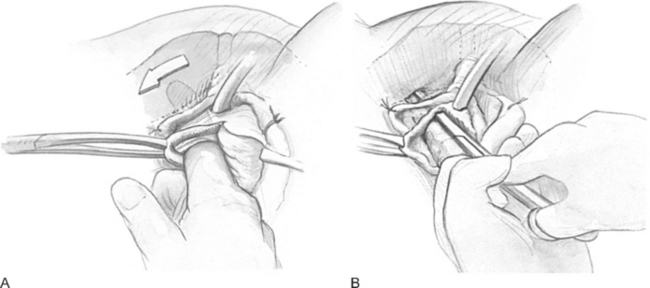
The TVT procedure was the first synthetic midurethral sling and was described by Petros and Ulmsten (1990), 1995). This ambulatory procedure was aimed at restoring the pubourethral ligament and suburethral vaginal hammock by using specially designed needles attached to a synthetic sling material. The synthetic sling material is made of polypropylene and is approximately 1 cm wide and 40 cm long. The sling material is attached to two stainless steel needles that are passed blindly from a vaginal incision made at the level of the midurethra through the retropubic space to exit at previously created stab wounds in the suprapubic area.


After this midurethral sling became well-established, techniques to surgically correct stress incontinences via a suprapubic approach were described (SPARC procedure [American Medical Systems, Minnetonka, MN]; percutaneous vaginal tape [PVT]). This involved passage of a needle from the suprapubic area to exit in a previously created incision in the vagina of the level of the midurethra. These procedures quickly became popular and were shown to be efficacious. Because they did require blind passage of the needle through the retropubic space, rare but serious complications were reported in the form of vascular and bowel injuries. These complications resulted in significant morbidity and rare cases of mortality.


Delorme (2001) reported on the first transobturator synthetic midurethral sling and described that the passage of the needle completely avoided the retropubic space and avoided any potential for serious complications in the form of vascular or bowel injuries as well as the potential for injury to the lower urinary tract. The initial description of this procedure involved the passage of specially designed needles from the obturator region, through the obturator membrane, around the ischiopubic ramus to exit in the open portion of the vagina. Shortly after the initial description, a procedure that involved passage of the needle from the vaginal incision to exit the obturator space was described.


TECHNIQUES OF SUBURETHRAL SLING PROCEDURES


Proximal Suburethral Sling and Modifications


The optimal route and surgical technique of a suburethral sling procedure placed beneath the proximal urethra and bladder neck must be considered preoperatively. Most surgeons perform a pubovaginal sling procedure via a combined abdominal and vaginal route, with the majority of the dissection being done vaginally. The procedure can also be performed entirely via abdominal approach, with tunneling of the sling between the urethra and the vagina. However, this approach increases the likelihood of urethral trauma and is currently rarely used. More recently, sling procedures have been performed through an entirely vaginal route through which the sling material is either anchored to the back of the pubic bone or sutured into Cooper’s ligament via a special needle ligature passage. The choice of sling material, approach, and operative technique is at the discretion of the surgeon. To date, no randomized trial has compared these various modifications and sling materials.


If the surgeon decides to use autologous tissue, it will most commonly be either fascia lata or rectus fascia. Harvesting of these tissues is usually performed before any vaginal dissection. The technique of harvesting fascia lata is determined by whether the surgeon prefers to do a complete pubovaginal sling, or whether a patch-type sling is performed. The fascia lata graft is usually approximately 4 × 6 cm in diameter. This can be obtained via a 3- to 4-cm transverse skin incision made approximately 8 cm above the midpatella, lateral to the knee and the lower thigh. Blunt dissection exposes the underlying fascial lata, and a piece of fascia is easily removed to serve as the patch for the sling procedure. Subcutaneous tissue is reapproximated, the skin is closed, and a pressure bandage is placed. If a full pubovaginal sling is used, a long piece of fascial lata can be obtained using a Wilson or Crawford fascial stripper. The technique for obtaining the full strip of fascia involves a blunt dissection of the fat away from the fascia lata all the way up the lateral side of the leg toward the greater trochanter. A 1-cm wide piece of fascia is usually removed using the fascial stripper. This will result in a 20-cm long piece of fascia. If a longer piece of fascia is desired, a second piece of fascia of a similar length can be obtained by repeating the same procedure. When this is performed, there should be a 1-cm wide area of fascia lata that remains between the two areas where the stripper has removed the tissue. In either case, closure of the fascia defect is not necessary.


The other autologous tissue that is commonly used for pubovaginal sling is the fascia of the anterior abdominal wall of the anterior rectus fascia. To obtain this fascia, a low transverse abdominal incision is made approximately 4 cm above the pubic symphysis. Blunt dissection is performed in the subcutaneous tissues until the rectus fascia is visualized. An appropriately sized piece of rectus fascia is harvested in the transverse direction using sharp dissection or electrocautery. The size of the strip is usually approximately 1 cm wide and 20 cm long. The fascial incision is closed using No. 0 delayed absorbable sutures, and the abdominal incision is packed until the vaginal portion of the procedure is completed.


The operative details for suburethral pubovaginal sling placed at the level of the proximal urethra are as follows:


4. The retropubic space is entered, using either blunt or sharp dissection (Fig. 16-2). Once the periurethral attachment of the urethra and the urogenital diaphragm has been penetrated on each side, one should be able to pass a finger along the backside of the pubic bone all the way to the inferior aspect of the rectus muscle.


Synthetic Retropubic Midurethral Slings


TVT


The patient is brought to the operating room and one of general, regional, or local anesthesia with sedation is administered. Broad-spectrum preoperative antibiotics, such as cephazolin, should be used. The patient is positioned in the dorsal lithotomy position. The authors prefer to use candy-cane stirrups for this. Patients should be in the horizontal or in slight Trendelenburg position to displace bowel away from the pelvis.


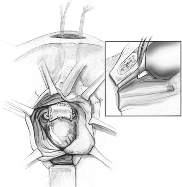
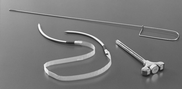
The instrumentation required for the procedure includes 30 mL of local anesthetic, such as lidocaine or Marcaine; a 22-gauge needle for infiltration; TVT kit (Fig. 16-5 [Ethicon Inc., Somerville, NJ]), which includes two 5-mm stainless steel needles connected by a 1.2-cm wide piece of polypropylene mesh; a nondisposable handle or introducer; an 18-French Foley catheter; catheter guide; and absorbable sutures to close the incisions.


Using a pen, the surgeon outlines the sites of the suprapubic stab incisions. These should be along the superior rim of the pubic bone, 2 fingerbreadths lateral to the midline, 0.5- to 1-cm long. The two suprapubic puncture sites should be anesthetized with local anesthesia. We prefer to use 1% lidocaine with epinephrine, 10 mL on each side. The injection should be taken down to the pubic bone to infiltrate the rectus fascia and muscle.


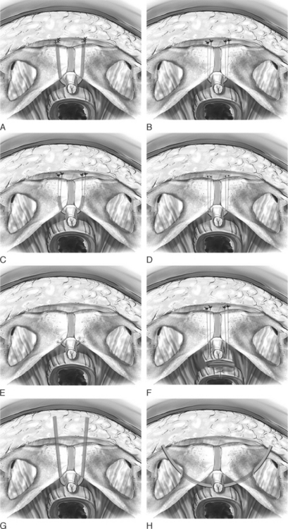
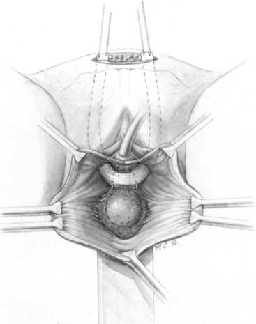
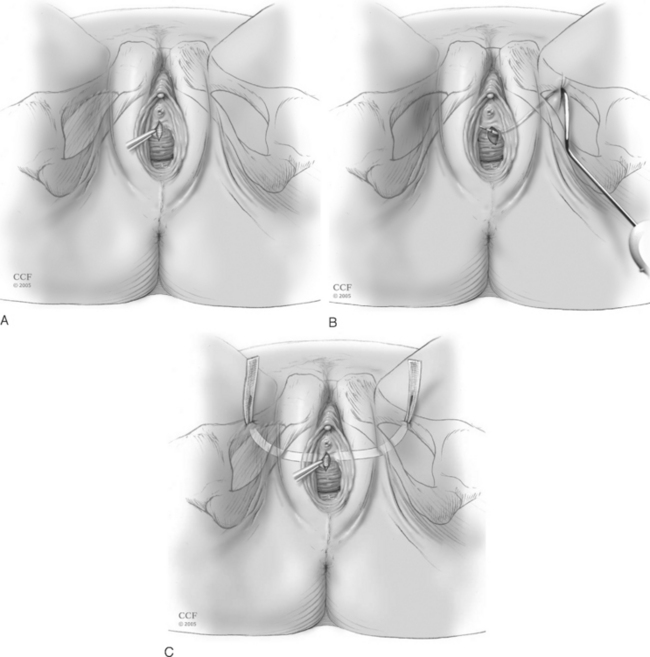
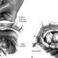
An 18-French Foley catheter should be placed and the bladder drained. A Sims or weighted speculum is placed in the vagina. Local anesthesia (8 to 10 mL) is infiltrated in the anterior vaginal wall to achieve hydrodissection and vasoconstriction. A 1.5-cm midline incision is then made at the midurethra, as shown in Figure 16-10, A. Palpating the Foley bulb at the bladder neck may be helpful to ensure that the incision is appropriately placed. With Allis clamps stabilizing the vaginal mucosa, the Metzenbaum scissors are used to dissect under the vaginal epithelium laterally to create a tunnel to the inferior pubic ramus. Unlike the bladder neck, at the midurethra the anterior vaginal wall and posterior urethra are fused and no natural plane of dissection is present. Hence, one must minimize creation of these tunnels to ensure that the tape stays in the proper position.


Once the tunnels have been prepared, the sling may be passed. Two stab incisions can be made in the suprapubic region over the marked areas. The bladder is completely drained, and the guidewire is passed down the Foley. The catheter guide is directed to the ipsilateral side of trocar placement to displace the urethra and bladder neck in the contralateral direction.

Stay updated, free articles. Join our Telegram channel

Mar 11, 2016 | Posted by in Reconstructive surgery | Comments Off on Sling Procedures for Stress Urinary Incontinence

Full access? Get Clinical Tree

Get Clinical Tree app for offline access