Skin graft

17 Skin graft


Skin grafting is a technique for the transfer of cutaneous tissue from one site of the body to another, often to cover large defects. Depending on the thickness of the dermis of graft that is harvested, skin grafts are defined as full-thickness or split-thickness.


Historical perspective


Skin is the largest human organ, covering 1.7 m2 in the average adult. It serves as an essential barrier with mechanical, immunological, and aesthetic functions. Skin transplantation has long fascinated humans and there is evidence that skin grafts were performed in India as early as 1500 bc for traumatic nasal amputation. Modern science has taught us much about the anatomy and physiology of skin.


Gaspare Tagliacozzi, one of the first reconstructive surgeons (1545–1599),1 described pedicled skin flaps from the arm for nasal reconstruction in patients while at the same time research was conducted by Giuseppe Baronio (1750–1811), who performed and published for the first time skin grafts in a lamb (Degli innesti animali, 1804).2 Jonathan Warren in Boston and Joseph Pancoast in Philadelphia were among the first to perform and describe autologous full-thickness skin grafting in humans using the arm as a donor site. Paul Bert, a French politician, showed in his thesis that skin graft survival is only possible if blood vessels of the recipient site are able to revascularize the graft. His work popularized this technique.3 For the techniques, Reverdin, a French student, described harvesting skin islands with the scalpel tip with transfer to a first web space wound.4 He discovered that these skin islands not only temporarily covered, but also actively stimulated, wound healing. Reverdin’s work was rapidly accepted and performed by multiple surgeons in the US. The term “dermoepidermoid” graft was introduced by Leopold Ollier (1830–1900), who also first used the split-thickness skin graft (STSG) to close entire wounds.5 He noticed that healing was achieved faster and wound contraction was minimized. The German surgeon Carl Thiersch (1822–1895) further developed the technique of skin grafting and introduced the concept of wound bed preparation by removing granulation tissue to achieve a clean wound bed to facilitate graft revascularization. The English ophthalmologist, John Reissberg Wolfe (1824–1904), from Glasgow, published the use of a full-thickness skin graft for ectropium correction.


With the development of skin harvest techniques the number of publications reporting clinical results of skin grafts increased exponentially. Later, James Carlton Tanner (1921–1996) revolutionized burn surgery by the introduction of meshing to expand skin grafts in order to cover larger wounds with minimal donor site morbidity.6


Reverdin’s technique was modified by John S Davis (1872–1946), who elevated the skin with a needle in 1914 to facilitate harvesting, a technique that he called “pinch graft.”4 In 1920 Ricardo Finochietto (1888–1962) developed a knife to elevate larger skin areas manually by controlling the thickness of the skin graft. Ten years later, the invention of the shave blade by Humby further facilitated skin harvest.


The introduction of the mechanical dermatome of Padgett-Hood and Reese in 1940 and 8 years later the development of the electric dermatome by Harry Brown significantly facilitated skin harvest of large surfaces in a controlled manner.


Anatomy and physiology


The skin represents approximately 8% of our total body weight, with a surface area of 1.2–2.2 m2. The skin is 0.5–4.0 mm thick and covers the entire external surface of the body, including the walls of the external acoustic meatus and the lateral tympanic membrane. The main function of skin is to protect body contents from the environment, including pathogens, temperature, and excessive water loss. Insulation, temperature regulation, sensation, immune function, and the synthesis of vitamin D are all critical functions of the skin. Skin loses regenerative capacity when lesioned down to the lower dermis and results in scar tissue when injured.


Epidermis


Skin has a complex three-dimensional structure characterized by two overlapping layers, the epidermis and the dermis. The epidermis, as the nervous system, derives after gastrulation from the neuroectoderm. Epidermis is the outer or upper layer of skin, which is a thin, semitransparent, water-impermeable tissue, consisting primarily of keratinocytes. These cells form a multilayered keratinized epithelium, similar to a wall of bricks. The basement membrane separates the epidermis from the dermal tissue and consists of a protein structure produced by basal keratinocytes. Basal keratinocytes are partially differentiated stem cells of the epidermis that provide the proliferative and regenerative capacity of the skin epithelium. The epithelium is metabolically active and continuously self-renews to maintain an efficient barrier function. Cellular homeostatic regulation is, as a consequence, very important: too little proliferation would bring a loss of barrier and excessive activity to hyperproliferative disorders, such as psoriasis. Homeostasis is granted by the basal epidermal cells, which periodically cycle, executing their program of terminal differentiation, a process that takes approximately 28 days. The differentiation of the keratinocytes is characterized by the progressive production of alpha-keratin, with migration towards the surface until the cells lose their intercellular connections (desmosomes), die, and become corneal lamina.


During this process, called cornification, basal keratinocytes produce tonofilaments (precursor of keratin) and then transform into the stratum spinosum as the desmosomes stretch the cells into spikes visible with a microscope. In the plasmalemma, the tonofilaments are connected to the desmosomes. Cells next start to produce keratohyalin, which aggregates in dense and basophilic granules, giving the name to the stratum granulosum. In these granules a histidine-rich protein, profilaggrin, becomes progressively filaggrin, which ultimately acts as a glue to keep keratin filaments together once the cells die and the cell membrane degrades.


As the cells divide and move up through the epidermis they eventually transform into the stratum corneum, a layer of dead cells, which ultimately is highly mechanically and chemically resistant due to chemical bonds between lipids and proteins. It is thought that cells die as the increasing proteins in their cytoplasm start to activate lysosomes. The stratum corneum provides an extremely effective barrier layer to keep water in and microorganisms out.


Also contained within the epidermis are melanocytes, Langerhans cells, Merkel cells, and sensitive nerves. Around 10% of the epidermal cells are represented by melanocytes, which derive from the neural crest. These complex dendritic cells produce melanin granules (contained in the melanosomes) that are then transported through dendrites into keratinocytes, providing color to the skin and protecting basal epithelial nuclei from ultraviolet damage. Melanocytes are anchored to the basal lamina by hemidesmosomes, but do not have desmosomic connections with other cells.


Langerhans cells are the immune cells of the epidermis and are important in generating a response to foreign agents, playing an important role in allograft rejection and contact dermatitis. These cells are situated in the stratum spinosum and, with long dendrites, slide between epithelial cells, without desmosomic connections to them. Langerhans cells share several features with macrophages of connective tissues.


Merkel cells are commonly found in the epidermis of palms, soles, nail beds, oral and genital areas. Merkel cells act as mechanoreceptors and thus are responsible for neurosensory transmission. These cells reside in the basal layer of the epidermis, often protruding into the dermal layer like nails. Merkel cells are connected by desmosomes to the neighboring cells.


Skin adnexal structures are epidermal derivatives that invaginate into the dermis with a lining of epithelial cells. They include hair follicles, sweat and sebaceous glands. These structures provide the basis for re-epithelialization following the harvest of an STSG.


Sensitive nerve supply to the skin is rich and extends through the basement membrane into the epidermis. Nerve fibers also go to skin adnexal organs that allow hair to become erect and sweat glands to secrete.


Blood vessel supply of the skin


Dermal vascularisation is particularly important, as blood vessels are not directly present in the relatively more metabolically active epidermal layer, glands, and hair follicles. Blood vessels set up a rich superficial plexus just underneath the basement membrane in the papillary dermis, facilitating nutrient transport to the epidermis. The blood vessels in the papillary dermis are arranged in the papillary plexus, with a rich network of capillaries in the papillae, which come in close contact with the epidermis. Deeper in the dermis is the reticular plexus from which small vessels distribute to the subcutaneous and deep dermal tissues to vascularize adnexal organs, including the hair follicle bulb. Arterial capillaries generate venous plexi, which have the same distribution of arterial vasculature. In the deep layers of dermis it is possible to find several arteriovenous anastomoses, particularly at the extremities (hands, feet, ears), where they exhibit strong muscular sphincters. The function of these structures is mainly under the control of the visceral nervous system, with the main function of thermoregulation and intravascular volume redistribution.


Blind-ended lymphatic structures are present in the dermis from where they connect to the reticular plexus and to larger vessels in the subcutaneous tissue. In this region, lymphatic vessels are larger, with valves, and drain in deeper lymphatic vessels called regional collectors. In the skin the lymphatic drainage is very active with multiple interconnections enabling lymphatic exchange. Circular skin and subcutaneous damage can therefore lead to problematic lymphatic stasis in extremities or in the genital area.


Stem cells and regeneration of skin


Basal epithelial keratinocytes are the committed stem cells of the epidermis. Constant self-renewal provides a new protective layer at the skin surface.712 Hair follicles contain multipotent stem cells that are activated upon the start of a new hair cycle and upon wounding to provide cells for hair follicle and epidermal regeneration. In the hair follicle stem cells reside in the bulge area. Bulge cells are relatively quiescent compared with other cells within the follicle.9,10 However, during the hair cycle, bulge cells are stimulated to exit the stem cell niche, proliferate,13 and differentiate to form the various cell types of the hair follicle.14 In addition, bulge cells can be recruited during wound healing to support re-epithelialization.15,16


The relative importance and exact contribution of bulge cells to wound healing are currently unknown as areas of the body such as the palms and soles that lack hair follicles still exhibit normal healing.


Hair follicles


Hair differentiates in a craniocaudal direction, 9 weeks after gestation, as mesenchymal cells populate the skin to form the dermis. Specialized cells in the dermal layer stimulate epithelial cells to proliferate and migrate downward into the epidermis, forming hair canals. The complete developed hair follicle contains an ectoderm-derived matrix and an underlying mesoderm-derived follicular papilla. There are three bulges that attach to the hair bulk. At the base the erector muscle develops, the middle part gives raise to the sebaceous gland, and the superficial bulge develops into an apocrine unit (Fig. 17.1).


image

Fig. 17.1 Skin grafts. (A) Split-thickness skin grafts (STSGs) are the preferred approach for the treatment of large superficial skin defects such as dermal burns. STSGs consist of the partial-thickness dermis and the epidermis. Depending on the thickness, STSGs are referred to as thin or thick STSGs. In thick STSGs superficial hair follicles can be included into the graft and restore hair growth and functional sweat glands in the future skin.


(B) Full-thickness skin grafts are limited in availability and are used in the reconstruction of aesthetic (face) or functional (hand) body areas. The graft is usually taken from behind the ear, or the inguinal or elbow fold. The graft consists of the complete dermal and epidermal layer, including hair follicles and glandular structures.


(C) Hair follicle: histological structures of the hair follicle. The presence of epidermal cells as far as the bulb explains the fast re-epithelialization after STSG harvest at the donor site. The infundibulum reaches from the entrance of the hair follicle into the skin to the apocrine gland. The zone between the apocrine gland and the sebaceous gland is referred to as the isthmus. The stem of the hair follicle is located between the sebaceous gland and the base of the erector muscle. The bulb is the deepest part of the hair follicle and contains the follicular matrix and papilla. This part grows and regenerates the hair after injury. If the bulb is lesioned the hair will not recover from injury.


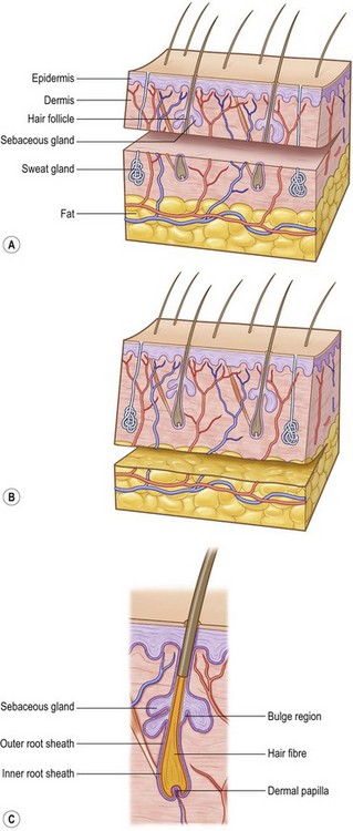
(Revised from Orgill, D. P. Excision and skin grafting of thermal burns. N Engl J Med 360: 893–901, 2009.62)


Histological structures of the hair follicle can be divided as follows. The part that reaches from the entrance of the hair follicle into the skin to the apocrine gland is the infundibulum. The zone between the apocrine gland and the sebaceous gland is referred to as isthmus. The stem of the hair follicle is located between the sebaceous gland and the base of the erector muscle. The bulb is the deepest part of the hair follicle and contains the follicular matrix and papilla. This part is growing and regenerates the hair after injury. If the bulb is lesioned the hair will not recover from injury.


Follicles can be found in different phases: anagen (proliferating phase), catagen (regression phase), and telogen (resting phase).


Although no new hair follicles are made postnatally, the lower portion of the hair follicle regenerates in order to produce new hair. Some of this capacity has been linked to the presence of multipotent epithelial stem cells. These cells can be found in the lowest permanent portion of the hair follicle – the bulge.13 Bulge stem cells are activated during the transition from telogen to anagen, to restart hair growth.


Glandular structures


Sabaceous glands are small saccular structures residing throughout the dermis, but are more common in thicker areas. These glands produce lipid-rich sebum on the surface of the skin and around the hair shaft. The function of sebum is still partially unknown but probably is linked to protection of the hair and contributes to the impermeabilization of the skin, giving protection from stings, parasites, and smell. Sebaceous glands are particularly large on the face, trunk, shoulders, and genital and perianal regions. When excessive quantities of sebum are produced – such as during puberty – the duct can be obstructed and ultimately bemay become infected or form cysts.


Sweat glands are divided into eccrine and apocrine glands. There are numerous eccrine glands in every region of the body except the tympanic membranes, lips, nail bed, nipples and clitoris. Their body has a glomerular structure and they excrete a clear odorless, hypotonic liquid. The secretion is stimulated mainly by an increase in body temperature, with the exception of some regions, such as palms, face, and axilla, where the main stimulus is emotional.


Apocrine glands are found exclusively in the axillar, perianal, periumbilical, areolar, preputial, scrotal, pubic and vulvar areas. While their structure is similar to that of the eccrine glands, these glands differ as regards the quality of their excretions, which are characterized by a thick milky, protein-rich fluid, which has a striking odor after bacterial colonization.


Science


Mechanisms of skin graft take


Skin grafting is the transfer of autologous skin cells left in anatomic order but without an intact blood supply. Therefore time and the recipient surrounding conditions limit the vitality. The operative procedure allows for nearly immediate coverage of large wound areas. Meshed grafts allow further expansion of skin but leave multiple small wounds that are re-epithelialized, mainly from the mesh within a few days. Skin can also be expanded through multiple small skin island grafts (as in Reverdin’s technique) that stimulate granulation tissue, probably by excreting growth factors. In STSGs, keratinocytes on the basal layer show high proliferation rates, which may ultimately stimulate growth factor excretion.17


Three phases of skin graft take are commonly described: (1) serum imbibition; (2) revascularization; and (3) maturation (Fig. 17.2).
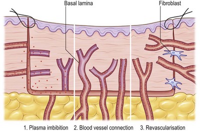


Serum imbibition


In the first days, before the graft revascularizes, oxygen and nutrients diffusing through the plasma between the graft and the wound bed will nourish the skin graft. Huebscher in 188818 and Goldmann in 189419 theorized that skin grafts might be nourished by host fluid before vascularization of the graft occurs. They referred to this as “plasmatic circulation.”18,19 Later, Converse et al.20 altered the term to “serum imbibition,” as fibrinogen changes into fibrin that fixes the skin graft on to the wound bed in the absence of real plasmatic flow. Converse’s studies show that skin grafts gain up to 40% of their initial weight within the first 24 hours after grafting and then this gain is reduced to 5% at 1 week postgrafting.20 In the first hours, passive absorption of serum from the wound bed causes edema, which resolves when the revascularization is functional (Fig. 17.2).


Revascularization


Revascularization is critical for long-term skin graft survival. Early studies in the 19th century2124 suggested a connection between the wound bed and graft vessels, referred to as inosculation,18,19,25,26 but the mechanism of revascularization remained unclear for many years.


Three hypotheses of revascularization are supported by the literature, each of them probably contributing to the process: anastomosis, neovascularization, and endothelial cell ingrowth. Anastomosis is the process of reconnection between the blood vessels in the recipient site wound bed and the graft.21,23,24,2729 Neovascularization is characterized by new vessel ingrowth from the recipient site into the skin graft. The last mechanism describes endothelial cell proliferation and sliding from the recipient site, utilizing pre-existing vascular basal lamina as a structure, while in the graft endothelial cells gradually degenerate.22,24,3032


The process of revascularization begins as early as 24–48 hours after grafting.33,34 Many authors describe vessel ingrowth mainly from the wound bed and less so from the wound margins, since no significant increase in blood vessels was seen in graft margins after skin grafting.21,24,3336 Studies supporting vessel ingrowth from the host as the main mechanism of skin graft revascularization have been controversial with respect to time course and the mechanism of host–graft vessel interactions.23,37 Some early studies demonstrated by intravenous injection with radioisotopes that blood flow in the graft was established 4 days after grafting.38 Similarly, studies using India ink showed graft vessel stain as early as 2 days postgrafting.23 More recently, it was demonstrated, using a transgenic tie2/pacZ mouse model, that vessel ingrowth appears in the periphery of the graft (following blood vessel regression in the graft) from day 3 until day 21.39 Zarem et al.22 suggested that the process of vascularization of full-thickness skin grafts in the mouse is dominated by vascular ingrowth from the recipient using a modified transparent skin chamber. Henry and Friedman37 proposed the theory that endothelial cells of superficial graft vessels degenerate and that host vessels profit from the basement membrane-covered infrastructure for new vessel ingrowth. In 1967, other investigators confirmed this theory using the graft hamster cheek pouch and showed a similar vessel pattern before and after grafting.40 Converse and Ballantyne further investigated endothelial cell ingrowth into the graft using diaphorase, a marker of viable vascular endothelium. They found increased diaphorase levels in the graft bed 4 days after grafting, supporting the theory of vascular ingrowth from the host. As very few functional anastomoses were present, the authors concluded that both mechanisms, inosculation and vascular ingrowth, were important in the process of revascularization.32 Vessel regression was also supported by NADH diaphorase activity loss during the first 4 days after grafting that was probably taken up by new vessel ingrowth.30,39,40 The conclusion that endothelial cells utilize preformed tunnels of basal lamina was triggered by the observation that initially empty graft vessels subsequently became infiltrated with leukocytes and that ingrowing vessels used the white blood cell-filled channel as a conduit.22 Later studies showed a central refilling of the graft vasculature as early as 48 hours, leading to the conclusion that early ansastomosis between host and graft vessels may play a major role in graft revascularization, as vessel growth takes about 5 µm/hour and angiogenesis would take at least 5 days to reach the 600-µm-thick murine dermis.22,41 In 1987 Demarchez et al.42 supported this hypothesis by grafting athymic mice with human STSGs. Double labeling of cross-reacting antifactor VIII and a human specific antitype IV collagen antibody showed initial anastomosis between graft and host vessels. Murine host cells gradually replaced vascular structure and the extracellular matrix of the skin graft. Later studies confirmed this hypothesis by observing a similar blood vessel network of the skin graft at the donor site and after revascularization of the graft between 96 and 120 hours after grafting.33


Capla et al. further showed that about 20% of blood vessels supporting the microcirculation on day 7 after grafting occurred by bone marrow-derived endothelial progenitor cell-derived vasculogenesis.39 Recent studies measured increased levels of growth factors related to hypoxia-induced angiogenesis (hypoxia-inducible factor-1 and vascular endothelial growth factor) 24–240 hours after grafting, a process that may also contribute to host vessel ingrowth (Fig. 17.2).33


Maturation


Once the skin graft is completely integrated, the same graft and surrounding tissues remodel and contract, similar to the last phase of wound healing after re-epithelialization is complete. Skin grafts take at least 1 year to complete maturation, with the extension of this process continuing for several years in burn victims and children. Scars from skin grafts can continue to improve for a number of years, often making prolonged conservative therapy worth considering.


Skin graft vascularization contributes to prevent underlying tissue contraction. Fibroblasts from surrounding tissues and from blood circulation become activated and repopulate the wound at the interface between the graft and the recipient site. As collagen is deposited, cross-linking allows the extracellular matrix to resist mechanical insults. Fibroblasts develop fibers called alpha-smooth-muscle actin (alpha-SMA) that exert contractile forces on the extracellular matrix. The development of alpha-SMA coincides with the differentiation of fibroblasts into myofibroblasts and wound contraction. During wound maturation, the epithelium from the edges of the wound produces a basal lamina on the open surface while sliding across and progressively covering the immature granulation tissue.


During the remodeling phase, all immature blood vessels necessary to support the initial phases regress and eventually disappear. The remodeling phase of wound healing is the longest, lasting from several months up to years.


Feb 21, 2016 | Posted by in General Surgery | Comments Off on Skin graft

Full access? Get Clinical Tree

Get Clinical Tree app for offline access