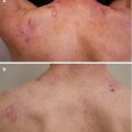
Figure 21.1
BIOCHIP™ mosaic after incubation with a bullous pemphigoid serum. Reactivity is seen with the dermal-epidermal junction of monkey esophagus and human salt-split skin as well as with recombinant BP180 NC16A directly coated on the BIOCHIPTM and BP230gC (C-terminal globular domain of BP230)-expressing HEK293 cells. No staining is observed with Desmoglein (Dsg) 1- and Dsg3-expressing HEK293 cells [9]
Which differential diagnosis can be made based on the BIOCHIP™ mosaic results and the case description?
1.
Epidermolysis bullosa acquisita
2.
Bullous pemphigoid
3.
Anti-p200/laminin γ1 pemphigoid
4.
Pemphigoid gestationis
5.
Drug eruption
Diagnosis
Bullous Pemphigoid
Case 2: A 46-Year-Old Female with Oral Erosions
A 46-year-old female was referred by her dentist with a 9-months history of erosions on the gingiva and right buccal mucosa. The lesions have been worsening during the course of the disease and led to a weight loss of 5 kg due to painful food intake. On examination in addition to the oral lesions, haemorrhagic crusts were found on the nasal mucosa and two crusted erosions on the chest and upper abdomen. Conjunctivae and genitalia were not involved.
Further workup included serological testing for autoimmune bullous disorders by BIOCHIP™ technology (Fig. 21.2).
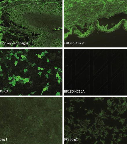
Figure 21.2
BIOCHIP™ mosaic after incubation with a pemphigus vulgaris serum. Intercellular labeling of the epithelium of monkey oesophagus and staining of Desmoglein (Dsg) 3-expressing HEK293 cells can be seen. No reactivity with the dermal-epidermal junction of salt-split human skin and the other substrates is observed [9]
Based on the case report and the BIOCHIP™ mosaic result, what diagnosis can be made?
1.
Pemphigus foliaceus
2.
Staphylococcal scaled skin syndrome
3.
Pemphigus vulgaris
4.
Oral herpes infection
5.
Mucous membrane pemphigoid
Diagnosis
Pemphigus Vulgaris
Discussion
In the group of autoimmune bullous disorders, the identification of target antigens enables physicians to differentiate between these frequently clinically similar appearing bullous diseases. Advances in the discovery of new antigens and the subsequent development of an increasing number of sensitive and specific assays for the detection of circulating autoantibodies allow the serological diagnosis in the majority of patients [1]. While the diagnostic gold standard is still the direct IF microscopy of a perilesional biopsy, in the majority of patients, diagnosis can be made by the combination of clinical findings and serology.
A step-wise analysis is usually employed starting with indirect IF microscopy using tissue sections (e.g. monkey esophagus for pemphigus and salt-split skin i.e. normal human skin in which the dermal-epidermal junction was separated by incubation in 1 M NaCl solution) followed by ELISA. At present, six ELISA systems based on the recombinant target antigens are available: desmoglein 1, desmoglein 3, BP180, BP230, type VII collagen, and envoplakin [2–8]. In some patients, additional analyses such as immunoblotting and immunoprecipitation are required to detect autoantibodies against e.g. laminin 332, the ectodomain of BP180, periplakin, and α2 macroglobulin-like 1. These assays are only available in specialized laboratories.
Related posts:

Stay updated, free articles. Join our Telegram channel

Full access? Get Clinical Tree


