Figure 19.1
Blisters of varying sizes on the limb
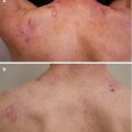
Figure 19.2 demonstrating an active blister with the background of an ulcer and secondary leucoderma
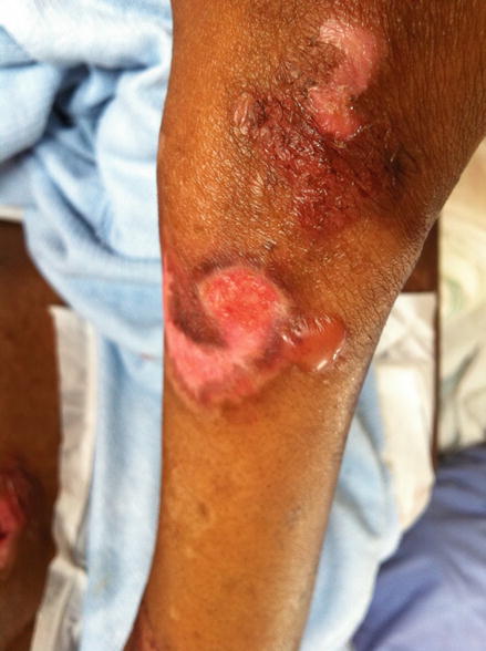
Figure 19.2
An active blister with the background of an ulcer and secondary leucoderma
What is your diagnosis?
EBA
Bullous Pemphigoid
Cicatricial Pemphigoid
Bullous SLE
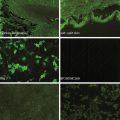
Biopsy showed a subepidermal blister with cell poor infiltrate and IFM was positive for IgG, C3 and IgM at the DEJ. She was found to have low complement C3 and ANF was positive in a titre of 1: 1250.
This patient was diagnosed with Bullous SLE and therapy with prednisone started at 0.5 mg/kg and Dapsone.
Bullous SLE
Bullous LE is an autoimmune blistering condition, often transient that occurs in the setting of systemic lupus erythematosus. It is commonly seen in young female patients of African descent [1–2].
Bullous lesions of Lupus Erythematosus can be single or widespread. They are commonly but not limited to sun exposed areas [1–2]. These lesions are painful but not pruritic. Patients with bullous lupus erythematosus meet the criteria for systemic lupus erythematosus hence (bullous SLE), but patient do exist with similar lesions but have fewer symptoms to meet the criteria (disease in evolution [1]). The occurrence of blisters is not related to flares of systemic disease [1]. Histology shows a sub-epidermal blister rich in neutrophils. Direct Immunofluorescence shows IgG, IgA, IgM and C3 in a granular or linear pattern at the basement membrane zone [1]. These antibodies target type VII collagen. Dapsone is first line therapy and is most effective [1–2].
Related posts:
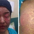
 An Elderly Patient with a Generalized Pruritic Eruption
A Healthy African Child with Blisters
An Elderly Patient with a Generalized Pruritic Eruption
A Healthy African Child with Blisters
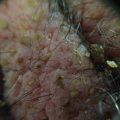 A 52 Year Old Man with Cerebriform Vegetating Masses on the Scalp
 Single Step Multivariant Analysis of Serum Autoantibodies in Autoimmune Blistering Diseases Using BIOCHIP® Mosaic Technology
 Detached Epidermis in an Adult Female
 A Man with a Blistering Rash
Stay updated, free articles. Join our Telegram channel

Full access? Get Clinical Tree


