Nasal septal deviation is a prevalent problem that can have significant quality of life ramifications. Septoplasty is commonly performed to provide qualitative and quantitative benefit to those with nasal obstruction owing to septal deviation. Although a standard, basic technique is often adequate for individuals with mild to moderate mid to posterior septal deviation, unique challenges arise with caudal septal deviation. Herein, multiple strategies that attempt to address anterior septal deviation are discussed. Anterior septal reconstruction has been shown to be a safe and effective means by which to address severe caudal septal deviation and long-term reduction in preoperative symptoms.
Key points
- •
Nasal septoplasty is one of the most common procedures in otolaryngology owing to the high prevalence of nasal obstruction from septal deviation.
- •
Traditional septoplasty is often inadequate in addressing nasal obstruction owing to anterior septal deviation, even when performed in conjunction with techniques to address nasal valve stenosis.
- •
Extracorporeal resection allows for correction of severe anterocaudal septal deviation but carries a risk of notching at the rhinion.
- •
Anterior septal reconstruction is a modified extracorporeal septoplasty technique that has been shown to address anterior septal deviation and minimize dorsal deformity and internal nasal valve collapse.
Introduction
Septal deviation is one of the most common causes of nasal obstruction and a prevalent problem among the general population. As such, septoplasty is among the most frequently performed procedures in otolaryngology and facial plastic surgery. This article provides a brief review of indications for septoplasty, surgical anatomy, and operative technique. We discuss basic and more advanced methods, with a special emphasis on advanced techniques, such as anterior septal reconstruction (ASR) for repair of anterior septal deviation.
Septal anatomy
A thorough understanding of the anatomy and function of the nasal septum and its surrounding structures is critical for surgical success. The nasal septum sits in the sagittal plane, extending from the maxillary crest inferiorly to the skull base superiorly and from the nasal tip anteriorly to the nasopharynx posteriorly and divides the nose into 2 nasal cavities.
The nasal septum is composed of both bony and cartilaginous components. Bony components include the maxillary crest, whose anterior extent forms the nasal spine, the perpendicular plate of the ethmoid bone, and the vomer ( Fig. 1 ). The quadrangular cartilage (QC) comprises the cartilaginous component and extends to the caudal-most aspect of the septum. Important landmarks of the QC include the anterior and posterior septal angles. Note that the former may extend beyond the nasal spine (see Fig. 1 ). The lateral aspects of the septal bones and cartilage are covered in mucoperiosteum and mucoperichondirum, respectively. At the junction of the bony and cartilaginous components lie decussating fibers, which are important to recognize and divide during surgery in maintain the appropriate subperiosteal and subperichondrial plane and prevent flap perforation.
The “keystone area” is the term that has been given to the attachment of the QC to the bony septum and nasal bones at the rhinion (see Fig. 1 ). Destabilization of this area can result in compromised dorsal integrity with saddle nose deformity. This complication is discussed in more detail below as one of the challenges of reconstruction of the severely deviated septum.
Important Surrounding Structures
Important surrounding structures that must be assessed and, if necessary, addressed during septoplasty include several bony and soft tissue structures. Perhaps the most important of these are the paired upper lateral cartilages, which attach to the cartilaginous septum dorsally. The angle between these and the septum forms the internal nasal valve usually measuring between 10° and 15°. This region is the most narrow point of the anterior nasal airway and is thus of high functional significance with regard to nasal obstruction. High septal deviations can result in narrowing of the internal valve.
The paired lower cartilages, whose medial crural attachments to the septum are 1 of 3 major tip support mechanisms. Note that release of the mucoperichondrium around the anterocaudal septum necessarily releases this attachment and deprojects the tip. Finally, the inferior turbinates are paired structures that help to regulate nasal airflow. When hypertrophied, they can contribute to nasal obstruction and are often reduced at the time of septoplasty. These structures are reviewed elsewhere in this volume and are not discussed further in this article.
Preoperative considerations
Surgical Indications
The most common indication for septoplasty is septal deviation with correlated symptomatic nasal obstruction. Note that a deviated septum alone (ie, without symptomatic obstruction) is not an indication for septoplasty. Other indications include improved access for endoscopic sinus surgery, lead point headaches, and source of graft (cartilage, bone, or mucosal) for patients undergoing skull base surgery.
Preoperative Workup
A thorough preoperative workup is essential for maximizing the potential for benefit and minimizing the risk of complication. Pertinent components of the patient interview include an analysis of symptomatic severity. In our practice, we routinely administer the Nasal Obstructive Symptom Evaluation (NOSE) or Rhinoplasty Health Inventory and Nasal Outcomes scale questionnaire. Patients with scaled NOSE scores of less than 30 are classified as “mild” obstruction and are unlikely to be operative candidates. A history of maxillofacial trauma or previous nasal surgery, autoimmune disease, drug use, bleeding disorders, use of anticoagulants, or systemic steroids is noted. Use of intranasal steroids is usually required by most insurance companies before surgical authorization in the United States at this time, and thus we recommend clearly documenting type of intransal steroid use, when used, and duration of use in the history.
Finally, it is of utmost importance that the surgeon determine if the patient currently or previously used vasoconstrictive nasal sprays or illicit drugs intranasally. There is some evidence to suggest that the use of these drugs can increase the risk of septal perforation. The senior author recommends discontinuance of the former for 6 months, and the latter for 1 year before surgery. Moreover, for the latter, we typically ask for a drug screen at the time of preoperative evaluation. Our experience is that patients are very eager to comply with these requests. Photodocumentation of nasal form with standard rhinoplasty views is recommended, although not required of septoplasty patients. If patients desire concomitant aesthetic surgery of the nose, a full rhinoplasty consultation ensues.
Preoperative Physical Examination
In addition to a full head and neck examination, anterior rhinoscopy with a nasal speculum is performed to visualize the nasal septum, as well as the nasal valves. Flexible or rigid nasal endoscopy can complement anterior rhinoscopy but, in this author’s experience, is not usually necessary. Mucosal status, as well as the size, of the turbinates are noted. Some key findings to note on examination are: (1) the location, degree, and direction of septal deviation (ie, parallel or perpendicular to plane of the septum), (2) Any posterior nasal spurs, which occur with abnormal bony growth under the posterior QC extension, (3) presence of septal perforation, (4) degree of inferior turbinate hypertrophy, (5) presence or absence of internal nasal valve stenosis (assessed with the modified Cottle maneuver), (6) any internal valve narrowing or lateral wall insufficiency which, if present is noted and graded, (7) the degree of tip support, and (8) any external deformities of the nose, especially dorsal and caudal deviations.
Surgical technique
Septoplasty can be performed either with a headlight and nasal speculum, or endoscopically. There are advantages and disadvantages to both techniques, but both have been shown to be successful means by which to correct septal deviation. Regardless of which approach is selected, the same basic surgical technique applies for traditional septoplasty, that is, that involving mild to moderate deviation of the mid or posterior septum.
Standard Endonasal Septoplasty Procedure Outlined
- •
The patient’s nose is first decongested with oxymetazoline in the preoperative holding area. After anesthesia is induced, 1% lidocaine with epinephrine is injected into the mucosa. In addition to providing local anesthetic and vasoconstriction, this injection, when done properly, can result in hydrodissection of the mucoperichondrium off of the septal cartilage, which facilitates flap elevation.
- •
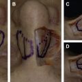
Next, using a nasal speculum for exposure, a 15-blade scalpel is used to incise the mucosa at the caudal septum down to the level of the cartilage. Either a Killian or hemitransfixion incision can be used; a hemitransfixion incision is more caudally positioned than a Killian incision and is preferred when caudal deviation is present ( Fig. 2 ).
Fig. 2
Incisions used in septoplasty. The hemitransfixion is more caudally positioned ( dotted line ) compared with the Killian incision ( solid line ).
- •
After making the incision, the senior author typically cross-hatches and scrapes the caudal QC to ensure the proper plane for flap development. After this, a sharp, curved elevator, such as a Cottle elevator, is used to elevate a mucoperichondrial flap. As this flap is raised broadly in the anterior to posterior direction, a blunt elevator such as a Freer elevator may be used. Note that, for standard septoplasty, dissection around the anterocaudal septum is usually not required. However, for caudal deviations, transition around the posterior septal angle is performed and the same plane dissected on the opposite side. If no external approach is planned, restoration of tip support with a septocolumellar stitch is required.
- •
Next, the surgeon must visualize and mark, if necessary, a 10- to 15-mm strut of cartilage dorsally and caudally. Two important issues must be mentioned. First, it is wise to preserve more than 10 mm (the senior authors prefers 15) at the keystone. In fact, if the septal deviation being treated is located inferiorly, the dorsal incision line of the septum is made must above it, preserving as much nondeviated septal cartilage as possible. Second, the posterior septal angle may exist anterior to the nasal spine (see Fig. 1 ), and the caudal strut must include 10 mm over the nasal spine. Thus, the caudal strut may be wider than 10 mm. Once the incision line is marked or visualized clearly, the QC is incised parallel to the dorsum from the keystone anteriorly and then angled inferiorly to define the new L-strut.
- •
A contralateral mucoperichondrial flap is then raised through this incision again first with a Cottle and then with a Freer elevator. After the bilateral flaps have been elevated, a determination is made regarding bony deviations (these are typically continuous with any QC deviations). The bony septum is incised carefully with a through-biting instrument with care taken not to (1) torque on the skull base or (2) release the keystone.
- •
The incised cartilage and bone is then removed en bloc using a noncutting instrument such as a Takahashi or bayonet forceps. Any remaining bony spurs can then be removed using a through-biting instrument, with care taken again to avoid torqueing the skull base or keystone support.
- •
Once all areas of septal deviation have been addressed adequately, the elevated flaps are laid back down. Care must be taken to assess for mucosal perforations and to ensure that, if present, these are not overlapping because this can result in permanent septal perforation. The senior author usually closes any linear perforations with fine chromic sutures. If opposing tears occurred, both sides are closed meticulously, and intervening tissue (such as morselized septal cartilage) is replaced between the flaps.
- •
Finally, the mucosal flaps are reapproximated and the mucosal incision is then closed. Surgeon preference dictates if, and what type, of packing or splinting is placed. The senior author performs a quilting suture and uses silastic splints in most septoplasty patients.
Related posts:


Stay updated, free articles. Join our Telegram channel

Full access? Get Clinical Tree


