This article presents a comprehensive review of past and present modalities in the surgical management of saddle nose deformities. Various surgical techniques, including allograft materials, are systematically reviewed. The senior author’s surgical experience and current management approach are highlighted.
Key points
- •
This article presents a comprehensive review of past and present modalities in the surgical management of saddle nose deformities.
- •
Various surgical techniques, including allograft materials, are systematically reviewed.
- •
The senior author’s (Hilger P) surgical experience and current management approach are highlighted.
Introduction
The saddle nose deformity constitutes one the most exacting anatomic, functional, and aesthetic challenges encountered by nasal reconstructive surgeons. Success dictates an approach focused on an understanding of the presenting etiology, precise anatomic analysis, and comprehensive surgical planning. Within a surgeon’s armamentarium, there has evolved a wide breadth of surgical options to effectively camouflage and reconstruct the saddle nose deformity. Operative management strives to achieve critical success in addressing the external aesthetic deformity and the inherent loss of functional nasal integrity. What follows is an in-depth review of these basic tenets in the operative intervention for the saddle nose deformity as well as the salient features in their technical execution.
Etiology and diagnostic considerations
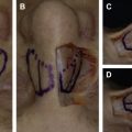
The principal anatomic deformity in the saddle deformity revolves around a deficit in nasal dorsal support secondary to the loss of septal cartilage and/or nasal bone height ( Fig. 1 ). The presenting etiology can be divided into 3 basic categories: iatrogenic, traumatic injury, and medical disease. Prior to the advent of nasal surgery, infectious causes, including septal abscess, syphilis, and leprosy, were the predominate disease entities. In the modern surgical era, operative sequela and traumatic injury constitute the vast majority of today’s saddle nose cases.
A minority of cases can, however, be linked to medical conditions whose nasal manifestations exhibit erosion of the dorsal septal cartilage and often degradation of other structural elements as well as crucial changes in the internal and external epithelial envelope. A differential diagnosis, including cocaine abuse, intranasal malignancy, and systemic inflammatory disease, warrant preoperative consideration. This latter group encompasses relapsing polychondritis, Crohn disease, sarcoidosis, lupus erythematous, and Wegener granulomatosis. In the vast majority of cases, a thorough screening history and focused physical examination, including anterior rhinoscopy, is sufficient to exclude an occult disease process. Resolution or, at minimum, stabilization of the metabolic disorders must be sought before surgical considerations.
Select patients with mucosal ulcerations, history of chronic sinusitis, or septal perforation can be considered for further investigation. Additional diagnostic modalities include nasal endoscopy, septal biopsy, and screening sinus CT scan or chest radiograph. Laboratory assessment in select cases includes erythrocyte sedimentation rate (ESR), C-reactive protein (CRP), angiotensin-converting enzyme (ACE), fluorescent treponemal antibody absorption, and antineutrophilic cytoplasmic antibodies (ANCAs). As to how extensive preoperative work-up should be, the sensitivity and overall clinical utility of nasal biopsies in cases of septal perforations and nasal ulcers are limited. Of the laboratory tests, ANCAs and ACE are more helpful than CRP and ESR in elucidating a systemic process among those patients with intranasal findings. Again, relying on a focused clinical history and physical examination more often than not excludes these rare entities. If chemical dependence is the genesis of the deformity, then prolonged sobriety must be the primary goal before surgical intervention. The authors have found the soft tissue contraction and vascular compromise provoked by cocaine abuse extremely challenging.
The clinical history should focus on prior nasal surgeries, trauma exposure, and nasal symptoms. For patients with a past history of nasal surgery and/or trauma, reviewing prior operation reports and CT imaging as well as preinjury photographs can be beneficial in gauging anatomic deficits, characterizing the premorbid state, and constructing a successful reconstruction sequence. Focused inquiry on symptoms of nasal obstruction enable both the surgeon and the patient to engage on the intricate link between form and function. From the perspective of both parties, it is especially important to delineate dynamic from fixed obstruction, anatomic from reactive pathology, and surgical benchmarks from medical management. As with any clinical endeavor with cosmetic implications, a patient’s sense of self and individual perception of the ideal nasal anesthetic assume center stage in the development of a successful treatment plan. Such success is born from a thorough clinical history during the initial consultation.
Physical examination and classification
The foundation of precise surgical management originates with a thorough physical examination, including both internal and external nasal components. Manual palpation of the nasal subunits yields significant information with regard to structural tip support, pliability of soft tissues, length of nasal bones, premaxillary support, and skin characteristics. Performing a Cottle maneuver, including its modified version, aids in establishing the role of the external and internal valve in nasal obstruction. It is helpful to evaluate the health of the nasal mucosa because some diseases demonstrate metaplasia of the respiratory epithelium with squamous cell replacement and resultant dry crusted debris accumulation. Decongestion of the nasal mucosa in the clinic permits delineation of dynamic obstruction and facilitates a complete examination of the nasal septum. As a principal dorsal support mechanism and a key contributor in the saddle presentation, the nasal septum with its associated mechanical strength, structural integrity, and contour needs to be fully assessed. Rhinomanometry and acoustic rhinometry represent useful adjuncts in quantifying nasal patency and may offer some predictive value in gauging surgical benefit with septoplasty alone when coupled with the physical examination. Rhinoscopy, cotton swab palpation, and nasal endoscopy constitute the primary tools for the surgeon in defining the contributions of the nasal septum to both dorsal and tip support as well as functional obstruction.
One of the core concepts in nasal reconstruction is evaluation of the skin soft tissue envelope (SSTE). As a fixed resource, the SSTE often serves as the limiting factor in how aggressive structural adjuncts might be placed into the nose. Housing the nasal bony and cartilaginous scaffold, the SSTE includes both the internal mucosal lining and the external skin. Manual palpation of the nasal tip and dorsum with digital manipulation of the internal nasal vestibule provides insight into skin tension, points of contracture, and pliability of the cartilage interface. Avenues to expand the SSTE may include skin grafting as well as local or composite grafting. Failure to incorporate the SSTE component in the reconstruction template, particularly in cases of revision surgery and prior trauma, frustrates the overall outcome.
In terms of stratifying the saddle nose deformity, there are several anatomic classification schemes in the literature. Integrating elements of progressive bony and cartilaginous deficits, these classifications systems can be helpful in establishing a rational approach to surgical intervention. One such system, offered by Daniel and Brenner in 2006, outlines a spectrum of saddle deformities ranging from mild depression of the dorsal cartilage to significant collapse of the bony vault ( Fig. 2 ). Because each individual nose may be funneled into a specific category, an anatomic centered approach in ascribing the severity of the nasal deformity has considerable merit. Accepting the premise that every nose is unique, the authors further submit that a tiered, structure-based evaluation aids surgeons in determining the right operation for the right nose. Focusing on the septum as the principal support mechanism provides surgeons a foundation to reconstitute dorsal projection, tip posture, and bony contour ( Fig. 3 ). Integrating these fundamental concepts with the preoperative physical examination and photographic analysis supplies the framework for precise surgical planning.
Patient counseling
With respect to preoperative counseling, only 14% of what is shared in verbal communication likely is retained. The skeletal and soft tissue distortions seen with these deformities require that patient and surgeon have a realistic image of what can be achieved with surgical intervention. Successful surgical outcomes for the saddle nose deformity often require more than 1 operation, with a reported minor revision rate ranging from 11% to 30%. Accepting the inherent variability in graft resorption, stable construct positioning, limited SSTE elasticity, and possible warping, it is important to educate patients on the complexity of the task at hand. It is the practice of the senior author to actively engage patients in a shared long-term perspective on achieving an optimal outcome. Moreover, in the authors’ experience, secondary surgical refinements are beneficial for approximately 30% of these patients. These should be candidly discussed with patients prior to the consideration of any surgery so that all involved in a patient’s care appreciate the complexity of these cases and refinement surgery. Combating the myopic view of a singular intervention with immediate permanency, patient counseling incorporates a frank conversation on the role and indications for minor revisions. This is understood to be part of the global care plan rather than a surgical failure of the initial operative plan. Even in the best of circumstances, the ultimate outcome is a compromise from the ideal and such expectations are essential if patient and surgeon are to be pleased. It is of particular import in cases of revision surgery, contracted SSTE, and bony vault reconstructions, where more than 1 consultation may be indicated. Establishing shared expectations between surgeon and patient is essential in moving forward from the day of surgery to when the cast comes off and, perhaps most importantly, the 12-month follow-up.
Surgical management
The efforts of Weir and Roe in late nineteenth-century New York state highlight the genesis of modern-day rhinoplasty. Valiant attempts to address the saddle deformity have incorporated a broad range of graft material ranging from ivory to gold to even the occasional sternum of a recently deceased duck ( Fig. 4 ). The grasp of nasal anatomy has matured in step with an empirical understanding of the limits in nasal augmentation material. Today’s reconstructive surgeons avail themselves of several options, including transient camouflage, autologous grafting, alloplastic implants, and homograft material. The fundamental challenge, however, remains not so much which material is used but rather the when, where, and rationale of how it is used. Assimilating the preoperative examination and the occasional intraoperative revelation, the reconstructive surgeon’s approach should incorporate an expectation and willingness to alter the surgical plan intraoperatively.
Referencing the tiered structural evaluation (see Fig. 3 ), surgical management is tailored to a 3-pronged graduated approach. Functional collapse of the dorsum is often associated with sacrifice of vertical nasal length, tip malposition, and a recessed columella. Stabilization and reinforcement of septal support serve as the foundation for correct tip positioning and dorsal augmentation. Progressively building support into these anatomically interdependent structures serves to enable contour augmentation and counteracts the inherent contractive forces of the SSTE ( Fig. 5 ) while correcting the airway impairment.
The nasal septum represents the cornerstone of dorsal support and needs to be formally addressed in surgical planning. Prior to focusing on the essential elements of tip posture and dorsal augmentation, the strength and mechanical support of the septum should be optimized. If there is a perforation amendable to repair, consideration for primary closure can be entertained in a combined or, rarely, a staged procedure. If the septum is intact, there is often a clear deficiency in the width or the height of the dorsal strut and caudal strut, respectively. The traditional septal L-strut of a 1.5-cm dorsal and caudal scaffold serves as a useful blueprint in the traditional nose, but the saddle nose is often depleted of septal resources and replete with structural laxity and scar. Addressing the septum in full, whether complete reconstruction with cartilage grafts, establishing a foundation in the premaxilla, or reinforcement with septal struts, sets the stage for subsequent tip and dorsal refinement. The premaxilla is an often overlooked concept and a significant contributor to the retraction seen at the columellar-labial angle ( Fig. 6 ).