, Shimin Chang2, Jian Lin3 and Dajiang Song1
(1)
Department of Orthopedic Surgery, Changzheng Hospital Second Military Medical University, Shanghai, China
(2)
Department of Orthopedic Surgery, Yangpu Hospital Tongji University School of Medicine, Shanghai, China
(3)
Department of Microsurgery, Xinhu Hospital Shanghai Jiao Tong University, Shanghai, China
In 1981, an international group of surgeons under the direction of Acland published their work on the “saphenous flap” [1–3].
21.1 Vascular Anatomy
The saphenous artery arises from the descending genicular artery. About 2 cm from its origin, the saphenous artery continues distally and pierces the roof of the adductor canal. After the artery passes through the roof of the adductor canal, it runs in the loose fascial space bounded by the sartorius muscle superficially, the adductor tendon posteriorly, and the bulging vastus medialis muscle anterolaterally.
It then courses straight down the leg in this space for 12–15 cm. In this part, it gives off early cutaneous branches that supply the large area of medial thigh skin above the knee. The cutaneous branches are given off 3–10 cm from the origin of the saphenous artery. The saphenous artery continues on its straight course, running from behind the tendon to enter the subcutaneous tissue. It supplies another large area of skin on the anterior and medial aspects of the leg below the knee (Fig. 21.1).
Fig. 21.1
Vascular anatomy of the saphenous artery
21.2 Illustrative Case
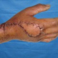
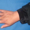
A 40-year-old man presented with severe scar ulcer on the popliteal space. Thirty-seven years before the patient suffered from traffic accident, early skin grafting was performed, but the ulcer occurred on the skin grafting area 5 years before (Fig. 21.2).
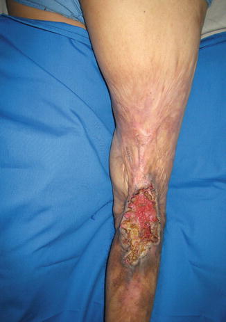
Fig. 21.2
Preoperative view
Flap Design
After radical debridement (Fig. 21.3), a pedicled saphenous perforator flap was designed. The key landmark for localizing the vascular pedicle is the sartorius muscle (Fig. 21.4). A line should be drawn from the anterior superior iliac spine to the medial epicondyle of the tibia, approximating the course of the muscle. The final position of the flap axis and skin paddle is determined after the position of the cutaneous perforators is confirmed (Fig. 21.5


Related posts:

Stay updated, free articles. Join our Telegram channel
Full access? Get Clinical Tree
