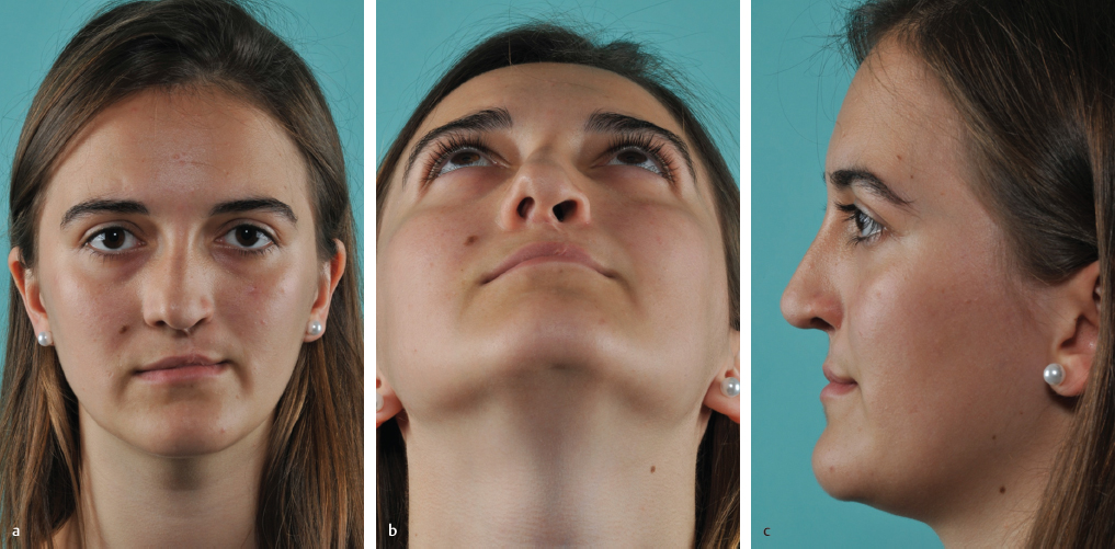
Chapter 13 13.1 Correction of Unilateral Cleft Lip Nasal Deformity 13.6 Septal Correction, Part 1 13.7 Septal Correction, Part 2 13.9 Psychology, Motivation, Personal Background In all cleft lip and palate (CLP) patients, a nasal deformity is part of the malformation and affects patients functionally as well as aesthetically. This complex deformity is due to a congenital anomaly and may partly be the result of previous surgical procedures. The nasal deformity in a unilateral cleft lip patient is totally different from that in a bilateral cleft lip patient. The nasal floor and the nostril sill are usually reconstructed during primary lip repair, which should be focused specifically on the repair of the divided oral ring muscle. The ala on the cleft side is repositioned symmetrically with the healthy side and sometimes the lower lateral cartilages (LLCs) also are fixed in a more symmetric way. Only a few surgeons try to position the dislocated anterior septum in the midline, which always follows the dislocated anterior nasal spine (ANS) to the noncleft side. New concepts attempt to reach early symmetry of the nostrils and the tip through subtle techniques without interfering with cartilage growth and avoiding the typical severe nose deformities that stigmatize these patients.1–3 According to Huffmann and Lierle,4 the classic cleft nose is characterized by 22 features, although not all of them are typically present in every patient. The external examination reveals a deviated nose to the healthy side due to the characteristic septal deformity. The anterior septum follows the dislocated ANS to the noncleft side. Therefore, the caudal border is always subluxated to the noncleft side. Very often, there is also an asymmetry of the nasal bones, so that not only the cartilaginous nose is deviated. Because of the malposition of the ANS, and thereby induced deformity of the caudal septum, there is always an oblique columella with asymmetric nostrils. In addition, the columella on the cleft side is shortened and the LLC (alar cartilage) on the cleft side presents an S-shaped distortion and is flattened and displaced laterally and/or cranially. Therefore, the nostril on the cleft side is horizontally orientated and presents as an oval shape in contrast to the physiological oblique orientation of the nostril on the noncleft side. The endonasal examination reveals a bowed septal deformity toward the cleft side, and the septum is deviated in all three planes—a so-called difficult septum being the result. Compensating for this typical deformity, the inferior turbinate of the noncleft side quite often shows a hypertrophy. The most lateral part of the lateral crus on the cleft side slants into the vestibule and forms the vestibular plica. The sill is often missing, depending on the effect of the alveolar bone grafting that is mostly performed at the age of 10 years. There might be a bony deficiency with a depression. In many patients with a unilateral CLP deformity, the correction of the nasal deformity has a high priority because it produces characteristic stigmata in the middle of the face. Upon meeting another person, usually you look into their eyes, but with a conspicuous nose like a cleft nose, your gaze will be drawn to it. This is realized and noticed by these patients, and it affects them. However, what worries them is not only the typical distortion of the nose, but also the functional impairment. Typically, there is a blockage in the cleft side and quite often breathing is blocked on both sides. The nose correction is, therefore, an essential part in the rehabilitation of CLP patients. The goal of surgery is straightening the deviated internal and external nose, creating symmetry of the nostrils, and giving a good contour to the tip. By correcting the anatomical deformities, good function can be achieved. In our concept, most of these severely deformed septums need an extracorporeal septal reconstruction, which means a temporary explantation of the whole septum.5 Because this drastically affects the growing zones, we only perform this kind of surgery 1 year after menarche in girls and 1 year after a change of voice in boys. Because of the complexity of surgery, we perform all cleft nose corrections under general anesthesia. There are five complex problems that need to be addressed: 1. The deformed septum. To straighten the nose, we need a straight septum. Because the septum presents mostly a three-dimensional deformity, in most cases we perform an extracorporeal septal reconstruction. 2. The displaced anterior nasal spine. Not only must the septum stay in the midline, but the malpositioned ANS also has to be fixed in its normal anatomical position. Therefore, in all major ANS displacements, we osteotomize the ANS, transfer it into the midline, and fix it there by microscrews and microplates. 3. The deviated nose. Because in most cases an asymmetric nasal pyramid is seen, we straighten the bony pyramid via parasagittal medial osteotomies as well as percutaneous lateral and transverse osteotomies.6 4. The distorted ala and deformed nasal tip. To reach symmetry of the nostrils, we generally perform a lateral crural steal technique on the cleft side and then replace the missing lateral crus with a cartilage graft. 5. The malposition of the alar base. Correction of the ala base is optional because this deformity depends a great deal on the previous surgery. There is a wide variety of typical ala base asymmetries, but in most cases the ala on the cleft side is too far lateral and too caudal. Using an open approach, we dissect the anterior septal angle, expose the caudal border, perform an extramucous dissection, and cut the upper lateral cartilage (ULC) from the septum. Then the upper and the lower tunnel of both sides are dissected. To remove the whole septum—if possible, take it out in one piece—a parasagittal medial osteotomy is performed. The author prefers a Lindemann fraise (Medicon Company, Tuttlingen) for that maneuver because an exact, straight bone cut can be achieved with this tool, while simultaneously removing some bone that otherwise might block the bone fragments during repositioning.6 To avoid any bad fractures, which might extend into the cribriform area, we make a bone cut downward from the bony dorsum with an angle of about 60 degrees. After dissecting out the base of the septum from the maxillary crest, we fracture the bony septum by pressing with a 5-mm chisel. It is important that the whole mucosal wall be freed from the septum before this step so that the mucosa will not tear during its removal. To achieve a straight neoseptum, or at least a straight L-shaped frame with the adequate dimensions, the length of the original dorsum and of the original caudal border have to be measured. Very often the explanted septum can be rotated 90 degrees so that the bony–cartilaginous junction becomes the new dorsum and the original dorsum gets the caudal septal border. Bent or weak parts are best splinted with thinned ethmoid bone. For reconstruction of the internal nasal valves and for keeping the neoseptum straight, we always apply spreader grafts. The use of spreader flaps is possible, but in these cases it is technically difficult. Thickened parts of the septum are always flattened with a cylindrical drill. Before replantation of the neoseptum, the ANS must be positioned in the midline. A side-to-side fixation of the replanted septum is only possible in cases of minor dislocation. Then the ANS is perforated with a drill to allow a transosseous fixation of the neoseptum placed next to the dislocated spine. In most cases, we cut the displaced ANS with a Lindemann fraise, put it in the midline, and fix it there by an angulated four-hole microplate that is secured by microscrews.6 The septum itself is sutured after replantation (see below) directly to the microplate. Before the septum is put back, the deviated and asymmetric bony pyramid has to be straightened. Parasagittal medial osteotomies are necessary for explanting the septum. The lateral as well as the transverse osteotomies are performed percutaneously. We do not respect Webster’s triangle, because in more than 10,000 rhinoplasties we have not seen problems with medialization of the lower turbinate’s head. After marking the low-to-low lateral osteotomy line, we make a stab incision, push away the vessels by scratching on the bone, and make a continuous transsection of the maxillary process with a 3-mm osteotome. The transverse osteotomy is performed analogously in the intercanthal line. After bringing the fragments in the right position, replantation of the neoseptum starts. Safe fixation of the replant is essential for success. After smoothing the dorsal line by trimming the nasal bones with a drill and the ULC with a scissor, the neoseptum is put back. To avoid the need of a columella strut, the neoseptum is put in a more caudal position to allow a tongue-and-groove technique for tip correction. First, the cartilaginous dorsum is reconstructed by fixing the neoseptum to the ULC with horizontal mattress sutures, followed by reconstruction of the keystone area. This is a most important step. The best and easiest way to fix the neoseptum to the nasal bones is a crisscross technique. After drilling a hole into the cranial–caudal part of the right nasal bone, a 4–0 PDS suture is placed downward and penetrates the upper edge of the LLC of the opposite left side. Then the nasal bone on the left side is perforated and the same thread is placed analogous through the right ULC—so that, in the end, the thread can get knotted on the outer side at the junction from the nasal bone to the ULC on the right side. In the end, the neoseptum is fixed to the ANS, respectively, to the microplate (see above). The length of the caudal border can be adapted by trimming the septum at its base. Then the anterior septum is fixed with three passes of a nonresorbable suture either through a drill hole in the ANS or directly to the microplate in case a transposition and osteosynthesis of the ANS is necessary. A strong and straight caudal border of the septum is also essential for tip correction. The goal is a symmetric cartilaginous framework of the LLCs. If trimming of the cephalic portion on the noncleft side seems appropriate, we do not resect this part, but dissect off the vestibular skin from the alar cartilage and turn the excess under it so that it gets stronger. Furthermore, this procedure helps to flatten the lateral crus. On the cleft side, we prefer to dissect out the lateral crus completely and form symmetric domes by a lateral crural steal technique. The complete dissection of the lateral crus enables us to give it a symmetric orientation to the noncleft side. By transferring the cartilage medially, a lateral deficiency may result, which can be corrected by a lateral crural strut graft or a batten graft taken from residual septum parts, straight parts from the concha, or from rib cartilage. For stabilizing the nasolabial angle and preventing postoperative drooping, we like a tongue-and-groove technique and fix the medial crura to the caudal border of the replanted septum. If it is not possible to follow this principle, we use a septal extension graft for the same purpose. If this does not work, a columella strut from a double-layered conchal graft is our favorite technique. The tip itself is contoured as usual with transdomal (interdomal) sutures and sometimes with additional intradomal sutures. The strong lateral crura can be molded by ala spanning sutures, which are combined with a tip suspension suture, fixing the tip complex to the dorsal septum (tip suspension with posterior sling technique). In case stronger lateral crura are needed to apply spanning sutures without any risk of creating an iatrogenic deformity, we prefer to use horizontal mattress sutures as suggested by Gruber7 for flattening and strengthening the alar cartilages. The soft tissue does not always follow the changes of the framework, and an asymmetry may persist. In such cases, we perform a triple-flap repair.5,8 The principle of this technique is to lengthen the short columella on the cleft side by a flap, based on the columella, and swing it inward 90 degrees after incising the vestibular skin for lengthening. The typically hanging ala on the cleft side is elevated into normal position by raising a second flap, based on the ala, and swinging inward accordingly. By transposing these two flaps, a gap results at the apex of the nostril. To achieve roundness, a third flap is created from the excessive vestibular skin, which remains after transposing the skin flaps. The position and the shape of the alar base on the cleft side depend a lot on the primary cleft closure and the effect of bone grafting. If there is a maxillary deficiency, we compensate it with diced cartilage fascia (DCF), made from allogenic fascia lata and autogenous diced ear or rib cartilage. We prefer rib cartilage because of its unlimited quantity. In minor asymmetries, we use fine-diced cartilage as free graft. In most cases, the ala on the cleft side is displaced laterally and/or superolaterally. If the alae are positioned at the same horizontal level, a lateral displacement can be corrected by an island flap from inside to outside, based on the small nasal muscles. This technique works inversely too and has the great advantage that, by the pull of the muscle pedicle, the effect of narrowing or widening is increased. Additionally, this lineament creates a nice crease and therefore gives a more natural appearance. If there is simultaneously a vertical asymmetry, we use transposition flaps for correction. If the ala base is positioned too cranially, we harvest a flap based on the upper lip mediocaudally to the ala. The lower incision has to be placed at the same level as the ala position on the healthy side. The vestibular skin is incised, the gap is filled with this transposition flap, and by closing the donor side, the ala is brought into a symmetric position. In case the ala base is placed too caudally, the same principle is used vice versa. An 18-year-old woman with left-sided CLP deformity presented for revision surgery after previous cleft nose correction (operation by Wolfgang Gubisch). The axis was deflected typically to the noncleft side, and the columella was oblique with consecutive asymmetric nostrils. The ANS was dislocated to the noncleft side and the caudal border of the septum was subluxated to the same side. The central septum was bowed to the left; on the right side, the lower turbinate was hypertrophic. The ala on the cleft side was pinched—the ala base was displaced superolaterally. In the profile, the maxilla was hypoplastic (the nasal tip was hanging) because the support to it was insufficient (Fig. 13.1a–f). After exposing the typical deformity using an external approach via an inverted-V transsection, we found the ANS dislocated 9 mm from the midline (Fig. 13.1g, h) to the noncleft side. The caudal border of the septum was displaced in the same way. After dissection of the deflected anterior septum (Fig. 13.1i) from the dislocated ANS, we cut the spine with the Lindemann fraise (Fig. 13.1j), put it in the midline, and fixed it there with an angulated four-hole microplate and micro screws (Fig. 13.1k, l). Before fixing the ANS, we augmented the hypoplastic maxilla with a DCF (Fig. 13.1m) from autogenous rib graft and allogenic fascia lata (Tutoplast; Tutogen Medical Gmbh). On top of the DCF, we placed a septal extension graft (Fig. 13.1n), harvested from the central septum. We fixed it to the anterior septum and then directly to the microplate (Fig. 13.1o), which secured the ANS in the midline. This septal extension graft kept the deformed septum straight. The hypertrophic turbinate bone on the right side was removed by a submucous resection. Tip asymmetry was balanced by suturing the medial crura to the septal extension graft using a tongue-and-groove technique (Fig. 13.1p) combined with a lateral crural steal technique (Fig. 13.1q). After trimming the asymmetric cephalic portions (Fig. 13.1r), the domes were contoured by intradomal sutures and then the tip was shaped by a transdomal suture (Fig. 13.1s). A very thin, extended shield graft was fabricated from the rib sutured in position and turned backward to cover the whole tip (Fig. 13.1t) to increase the projection. To avoid any irregularities, this construction was covered with a single layer of allogenic fascia lata (Tutoplast) (Fig. 13.1u). Before putting the skin flap back, an underbatten graft was placed to the right ala (Fig. 13.1v), fixed with transcutaneous sutures (Fig. 13.1w), and a rim graft was placed to the right side. After closing the columella, before closing the infracartilaginous incision, the final contouring was performed with a finely diced, paste-like cartilage (Fig. 13.1x), using it as a free graft injected through a tuberculin syringe (Fig. 13.1y).
Rhinoplasty after Cleft Lip Repair
13 Rhinoplasty after Cleft Lip Repair
13.1 Correction of Unilateral Cleft Lip Nasal Deformity
13.2 Surgical Anatomy
13.3 Indication
13.4 Surgical Principles
13.5 Operative Technique
13.6 Septal Correction, Part 1
13.6.1 Correction of the ANS
13.6.2 Correction of the Bony Pyramid
13.7 Septal Correction, Part 2
13.7.1 Tip Correction
13.7.2 Correction of the Alar Base
13.8 Case Examples
13.8.1 Case 1
Plastic Surgery Key
Fastest Plastic Surgery & Dermatology Insight Engine