Reduction rhinoplasty techniques include maneuvers that weaken the nasal osseocartilaginous framework. The structurally compromised anatomy remaining after reductive surgery may be left with inadequate strength to withstand postoperative contractile forces. Significant aesthetic and functional deformities requiring revision rhinoplasty may develop. This article reviews common causes of nasal obstruction after primary rhinoplasty. The discussion of etiology is based on both the anatomic description of nasal subsites (middle vault and lateral walls) as well as an explanation of why certain techniques lead to functional problems in these areas. Revision rhinoplasty techniques for correcting these problems are discussed in detail.
Key points
- •
A smooth, symmetric, well-proportioned surface contour with a stable underlying structural foundation is the ultimate aesthetic goal for rhinoplasty.
- •
The relationship between the upper and lower lateral cartilages and the septum is crucial in maintaining nasal tip and dorsal support.
- •
Secondary nasal obstruction is commonly caused by inward malposition of the upper lateral cartilages and internal valve area and/or collapse of the lateral crura and external valve incurred during reductive primary surgery.
- •
Correction of middle vault collapse may be accomplished through spreader grafts that may be extended in a cephalic, caudal, or dorsal direction depending on anatomic need.
- •
Correction of lateral wall collapse is effectively treated with lateral crural strut grafts (LCSGs), which may have variable length, stiffness, and position depending on degree of pathology.
Introduction
Rhinoplasty has been described as one of the most challenging procedures in plastic surgery, with revision rates reported as high as 15% to 20% after primary procedures. Many rhinoplasty techniques rely on resection of the nasal osseocartilaginous framework to achieve aesthetic objectives. The weakened nasal framework remaining after reductive surgery may have inadequate strength to withstand the contractile forces of healing. Patients often present with significant aesthetic and functional deformities requiring revision surgery.
The unfavorable interplay of soft tissue contracture and a weakened structural framework can lead to severe obstruction and cosmetic deformity. In revision rhinoplasty, the surgeon may need to build new constructs out of transplanted material that mimics the shape and function of the original anatomy. Thus, when revision rhinoplasty aims to rebuild a nose that has been over-reduced and structurally compromised, the task is incredibly difficult.
There are several categories of rhinoplasty complication ( Table 1 ). In cases of postrhinoplasty nasal obstruction, the most likely causes are destabilization of nasal structures and/or excessive reduction or excision of the nasal framework. The most common areas in which these problems occur are in the middle vault and lateral walls of the nose. In the middle vault, cartilaginous hump reduction, especially when accompanied by narrowing osteotomies, leads to inferomedial migration of the upper lateral cartilages and internal valve compromise. In the lower nose, narrowing tip techniques lead to medialization of the lateral crura and alar and external valve collapse.
| Minor (error of technique) |
|
|
| Error of omission (failure to execute a needed step) |
|
|
| Failure to restabilize structures that have been weakened during surgery |
|
|
| Excessive excision (overaggressive reduction of the nose) |
|
|
| Gross error of judgment |
|
|
Correction of these deformities is best treated with structural grafting techniques that strengthen the compromised anatomic structures and restore their stable relationships to one another.
Preoperative assessment
The consultation between a patient seeking a revision rhinoplasty and a surgeon is crucial to understanding motivations and establishing common goals. The magnitude of functional impairment should be determined. Utilization of the Nasal Obstruction Symptom Evaluation (NOSE) instrument can help quantify the degree of obstruction and also track improvement and long-term function after revision. Patients must understand that revision surgery is more difficult, with potentially limited outcomes given the degree of trauma incurred previously. Patients with previous reduction rhinoplasty and functional impairment should understand that certain areas of the nose may need to be expanded to allow for restored function. Use of digital imaging to render such visual outcomes may be helpful in these cases. Surgery should only be undertaken if both patient and surgeon have reached common and realistic goals.
When evaluating the revision rhinoplasty patient, it is important to determine what surgery has been done in the past, along with the timing and number of previous procedures. Old operative notes are sometimes helpful but should never be taken at face value, because they may not contain all details of the previous procedure. The external nasal contour and the internal airway must be assessed to diagnose and localize the deformities.
An accurate assessment of the aesthetic and functional problems helps surgeons formulate a surgical plan and select the appropriate techniques.
The physical examination should be conducted in a systemic fashion. The symmetry and width of the nose should be assessed on frontal view, including evaluation of the brow-tip aesthetic line, which can help to identify middle vault deformities, such as middle vault collapse or an inverted-V deformity. These features may reveal an underlying middle vault and internal valve compromise. Assessment of the lower third of the nose is important to assess lateral wall stability and contour. Findings, such as dynamic inspiratory collapse, alar retraction, supra-alar pinching, alar narrowing, and bossae, may be evidence of previous destructive tip work. Both inspection and palpation of the nasal septum should be performed to determine abundance, stability, and position of the nasal septum.
Middle vault
In normal nasal anatomy, the middle vault consists of the paired upper lateral cartilages and their associated internal nasal mucosa. The upper lateral cartilages support the internal valve and determine the external contour of the middle third of the nose. The upper lateral cartilages attach cephalically with the nasal bones and medially with the cartilaginous septum. At their upper aspect, the upper lateral cartilages display a dome-shaped cross-sectional geometry, conforming the shape of the nasal bones as they emerge from the undersurface of the bones. Here, the upper lateral cartilages fuse to one another horizontally across the dorsum. Caudally, the upper lateral cartilages take on a more triangular cross-sectional geometry. In this region, they are associated with the paired lower lateral cartilages forming the scroll region. The internal nasal valve area is defined as the connection between the caudal margin of the upper lateral cartilage, the head of the inferior turbinate, the floor of the nose, and the septum. Often in an unoperated nose, this is naturally the narrowest point of airflow and area of greatest flow resistance.
The most common obstructive problem seen in the middle vault in revision rhinoplasty is malposition of the upper lateral cartilages in a medial, inferior, and/or ventral direction. This is often due to collapse of the upper lateral cartilage onto the dorsal septum after cartilaginous hump reduction, especially when the dorsal margin of the upper lateral cartilages is resected en bloc with the dorsal margin of the septum. This maneuver greatly reduces the dorsal-ventral dimension of the upper lateral cartilages, leading to the characteristic inferomedial malposition along with the associated middle vault pinching, characterized by deep vertical shadows flanking the dorsum. These problems may also involve an inverted-V deformity in which the caudal aspect of the nasal bones are skeletonized and visibly revealed due to the upper lateral cartilages pulling away from the bones inward. The three-fourths view is particularly helpful in assessing middle vault and internal valve 3-D deficiency, because the upper lateral cartilage depression is seen as a contour depression from this oblique angle. Patients with short nasal bones and long upper lateral cartilages are especially prone to these problems. Aggressive narrowing osteotomies can exacerbate the above middle vault problems by causing additional medialization of the upper lateral cartilages, by virtue of their connection to the nasal bones.
There are several strategies that may be used to prevent these problems during primary rhinoplasty. First, the amount of hump reduction may be minimized by preserving dorsal height. Adding radix volume and/or increasing tip projection in certain cases may allow for less dorsal reduction while still resulting in a straighter dorsum. Second, preserving the dorsal-ventral length of the upper lateral cartilages may reduce the risk of inward collapse of the upper lateral cartilages after hump reduction. This requires that component reduction of the hump be performed, in which the upper lateral cartilages are first separated from the dorsal septum and the septum lowered first. Preserving upper lateral cartilage dorsal height may then allow for inward folding of the upper lateral cartilages (spreader flap or autospreader) to maintain middle vault stability and function. Third, spreader grafts can be used to restore or precisely modulate middle vault and internal valve width and support.
In cases of revision surgery for the over-reduced nose, the spreader grafting technique is most helpful. There is usually a deficiency of native upper lateral cartilage left for in-folding and the dorsal septum has been overly lowered. It is the preference of the senior author (Kim DW) to approach this correction through an open rhinoplasty approach with an anterior dissection to the dorsal septum, typically first by establishing a submucoperichondrial plane at the anterior septal angle. This dissection must be executed meticulously because prior caudal septal truncation and septal harvest may have distorted the anatomic landmarks and tissue planes. If a weak, compromised L-strut is encountered, a decision must be made as to whether or not the L-strut is strong enough to serve as a foundation for the subsequent spreader grafts (and, if needed, tip work). If L-strut reconstruction is needed, this should be done at this point prior to any work on the middle vault or tip.
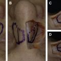
At this point, the surgeon must determine the appropriate dimensions of the spreader grafts. The frontal width should be assessed by using both internal and external landmarks. Internally, the cephalic aspect of the spreader grafts should be wide enough to provide a smooth transition to bony dorsal aesthetic line. Ideally, the graft allows for a smooth transition between the bony vault in 3-D. This is best checked by assessing continuity with the skin envelope down after spreader graft placement from several perspectives (frontal, lateral, and all angles between). If there is asymmetry of the dorsal septum, asymmetric width of the spreader grafts can help camouflage this. Caudally, the spreader graft width should taper as it approaches the anterior septal angle but remain wide enough to support and open the internal valve. In many cases, multiple layered spreader grafts may be needed to provide enough width for any given revision nose ( Fig. 1 ).