Mild
PaO2/FiO2 ratio from 200 to 300 mmHg with PEEP or CPAP ≥ 5 cmH2O
Moderate
PaO2/FiO2 ratio from 100 to 200 mmHg with PEEP ≥ 5 cmH2O
Severe
PaO2/FiO2 ratio < 100 mmHg with PEEP ≥ 5 cmH2O
The use of LTV in patients with burn injury has been extrapolated from the ARDSNet study, which is controversial due to the fact that this trial excluded patients with burn injury [12]. A single center, randomized controlled trial was performed on 62 patients with burn injury that randomized them to either LTV or mechanical ventilation with a high frequency percussive ventilator [8]. Baseline demographics, percentage of total body surface area (TBSA) burned, and rates of inhalation injury were similar in both groups. Nearly one-third of patients in the LTV group failed to meet pre-determined goals for oxygenation and ventilation and had to be placed on a rescue mode of mechanical ventilation. Two thirds of patients with smoke inhalation failed LTV. Burn injury and the loss of chest wall compliance due to edema or burn eschar, the hyper-catabolic state associated with burns and other variables make LTV a less effective treatment strategy.
Therapies for Refractory Hypoxemia in Patients with ARDS. Severe hypoxia induces vasoconstriction of the pulmonary vasculature which in turn causes pulmonary hypertension [13]. It is hypothesized that vasodilation of blood vessels perfusing aerated lung tissue with inhaled pulmonary vasodilators would redistribute blood from less ventilated regions of lung, reducing shunt fraction and correcting pulmonary hypertension. This process should then improve oxygenation and decrease mortality; however, it has not proven to be the case in randomized clinical trials [14]. Meta-analysis of the data from randomized trials reveals that while inhaled nitric oxide will improve oxygenation it does not decrease mortality and may increase rates of AKI [15]. The role of inhaled nitric oxide as a therapy for refractory hypoxemia is unclear.
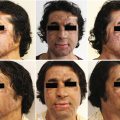
Prone positioning is a viable treatment for refractory hypoxemia in adult burn-injured patients with severe ARDS. In a trial involving 18 burn ICU patients, the average PaO2 to FiO2 ratio increased from an average value of 87 prior to prone positioning to 236 at 36 hours after initiating prone positioning [16]. While facial ulcers developed on 4 patients, there were no unintentional extubations.
Extracorporeal membrane oxygenation (ECMO) technology has improved in the last decade with respect to its use in burn-injured patients [17]. In our burn center, the survival to hospital discharge rate for patients with severe ARDS due to burn injury, inhalation injury, or toxic epidermal necrolysis treated with ECMO is 57% [18]. This is a viable treatment strategy to reduce mortality in properly selected patients.
19.4 Cardiovascular
Burn injury can disturb multiple physiologic variables that affect the function and performance of the circulatory system. Volume loss into burned and non-burned tissue results in intravascular volume depletion which reduces the venous return to the heart, resulting in decreased cardiac output. Large TBSA burns induce myocardial depression which can cause a global decrease in contractility and a reduced left ventricular ejection fraction. This myocardial depression is pronounced during the initial presentation and this is usually followed by a hyper-dynamic phase in which cardiac output is augmented by an increase in heart rate and a reduction in afterload caused by vasodilation which is mediated by inflammatory cytokines. When patients with burn injury develop hypotension, clinicians will usually attribute this to volume depletion and vasodilation, and it is often the case early in the patient’s resuscitation. Clinicians should put forth effort to make an accurate determination of the patient’s intravascular volume status as unnecessary fluids or fluid creep can cause edema to form in the brain, lungs, bowel wall, renal parenchyma, skin and soft tissue. Determining intravascular volume status using the physical exam, vital signs, intake and output flow sheets, urine output, and calculating insensible losses is difficult to do reliably and in a way that is easy to reproduce among members of a care team. Any one, or many, of these parameters can be confounded by factors other than intravascular volume and this assessment relies upon multiple points of data rather than any one gold standard measure.
Right sided or static measures of hemodynamics and cardiovascular performance, namely the central venous pressure and pulmonary artery occlusion pressure have been demonstrated to be inaccurate in healthy patients, let alone those that are critically ill [19]. For this reason, dynamic measures of cardiac performance such as using an arterial line to calculate arterial wave form pulse pressure variation or stroke volume variation, cardiac output and cardiac index or using beside transthoracic or transesophageal echocardiography in real time is preferred.
When an intravascular volume assessment reveals that the patient has been adequately resuscitated and the patient remains hypotensive with a mean arterial pressure less than 65 mmHg, vasopressor therapy should be initiated. While there are no large, multi-center randomized controlled trials on this subject, our practice is to initiate vasopressin first followed by norepinephrine, epinephrine, and phenylephrine. The use of vasopressin in burn patients already on norepinephrine had a norepinephrine sparing effect and resulted in graft loss in only one of 30 patients involved in the study [20]. In patients with echocardiographic evidence of myocardial depression and left ventricular systolic dysfunction, clinicians should consider the use of dobutamine. Additionally, if patients have high vasopressor requirements, especially in the setting of septic shock, clinicians should consider initiating stress dose corticosteroids (hydrocortisone 100 mg IV every 8 h) for relative adrenal insufficiency associated with sepsis. [21].
19.5 Renal
KDIGO staging criteria for patients with AKI
Creatinine criteria | Urine output criteria | |
|---|---|---|
Stage 1 | ↑ 0.3 mg/dL in 48 h or by >50% over 7 days | <0.5 mL/kg/h for >6 h |
Stage 2 | ↑ Creatinine of >100% | <0.5 mL/kg/h for >12 h |
Stage 3 | ↑ Creatinine of >200% | <0.3 mL/kg/h for ≥24 h or anuria for ≥12 h |
Traditional indications for initiating renal replacement therapy
Complication | Criteria for initiating renal replacement therapy |
|---|---|
Metabolic acidosis | Refractory to sodium bicarbonate infusion |
Electrolyte imbalance | Hyperkalemia refractory to medical management |
Toxic ingestion | Small molecular size and low level protein binding so as to be cleared by dialysis |
Uremia | Uremic encephalopathy, pericarditis, gastropathy, or bleeding due to uremic platelets |
Intravascular volume overload | Pulmonary edema refractory to diuretic therapy |
The literature demonstrates that early and intensive RRT may provide the best benefit for critically ill patients, particularly in those with AKI diagnosed early in their clinical course. Critically ill burn patients who develop AKI have mortality rates that are over 20% higher than the general critical care population [23, 24]. Historically, these patients were often not considered candidates for intermittent hemodialysis (IHD) due their hemodynamic instability by the time they developed a traditional indication for RRT. Continuous veno-venous hemofiltration (CVVH) is known to be well tolerated in the setting of shock. In a recent study, patients with greater than 40% total body surface area burn and AKIN stage 3 AKI or AKIN stage 2 AKI with shock were started on CVVH, even if they did not meet traditional criteria for initiating RRT. When these patients were compared to historical controls, they had a 24% lower in-hospital mortality rate, a 33% lower 28-day mortality rate, a dramatic reduction in vasopressor requirement and an average PaO2/FiO2 ratio increase of 153 within 24 h [25]. What mode of renal replacement therapy will provide maximal benefit to the patient, at what dose and when therapy should be initiated are questions that investigators have sought to answer with the best data available. We review these controversies below.
Mode. In a review of nine randomized trials comparing outcomes in patients placed on intermittent compared to continuous therapy, there was no standardization of the timing, criteria for initiation or dose of renal replacement therapy [26]. This review concluded that there was no statistically significant evidence that the initial RRT modality influenced mortality or recovery of renal function. There was a trend observed that CRRT was associated with less hemodynamic instability and better control of the patient’s fluid balance. Another meta-analysis reviewed 23 randomized controlled trials and 16 observational studies to investigate if there was any difference in rates of renal recovery among severe AKI survivors [27]. Pooled analysis of the RCT’s did not reveal a statistically significant difference in renal recovery based on RRT modality used. A pooled analysis of the observational studies did demonstrate a higher rate of renal recovery among patients receiving continuous therapy.
The main factor to be considered in deciding to use intermittent or continuous RRT is the patient’s hemodynamic stability. Advanced age, female gender, diabetes, low blood pressure prior to dialysis, hypoalbuminemia, and higher BMI are all associated with increased risk for intradialytic hypotension [28]. If removal of intravascular volume exceeds the patient’s ability to refill their plasma volume, then hypotension will occur. The level of sodium in the dialysate, plasma albumin levels and hydrostatic capillary force all influence plasma refill [29]. The patient’s osmolality declines during dialysis due to the rapid removal of urea and shifts in plasma sodium concentrations which will lead to slower plasma refill and eventually hypotension [29]. For patients who are hemodynamically unstable or who cannot tolerate large volume shifts and rapid changes in plasma osmolality, continuous modalities offer the benefit of renal replacement therapy with less risk of hypotension. Patients with neuro-trauma or other conditions that elevate intra-cranial pressure cannot tolerate the osmotic shifts that occur during IHD and it is contraindicated in this patient population [30].
Dose of Renal Replacement Therapy. The currently recommended dose for renal replacement therapy is 20–25 mL/kg/h [30]. Most patients do not have day after day of uninterrupted CRRT. Therapy can be interrupted by trips to radiology for imaging studies or dysfunction with the circuit caused by clotting or malfunction of the dialysis access line. In one study, interruptions in RRT ranged from 8 to 28% of the total treatment time [31]. Clinicians should target a prescription of 25–30 mL/kg/h and above so as to account for interruptions in therapy and insure that the patient is receiving the minimal delivered dose of 20–25 mL/kg/h. The current recommended dose is based on several clinical trials that compared different doses of renal replacement therapy and their impact on clinical outcomes. The VA/NIH Acute Renal Failure Trial Network randomized 1124 patients with acute kidney injury and failure of at least one non-renal organ or sepsis to intensive therapy (CRRT dose of 35 mL/kg/h) or less intensive therapy (CRRT dose of 20 mL/kg/h). The primary end point was all-cause mortality by day 60. There was no difference in the rate of all-cause mortality, or in duration of renal replacement therapy, or the rate of renal recovery [32]. The RENAL Replacement Therapy Study Investigators conducted a multi-center, randomized controlled trial to compare the effect of different doses of renal replacement therapy on 90-day mortality. They randomized patients with AKI and critical illness to post-dilution CVVH with a dose of 40 or 25 mL/kg/h. There was no statistically significant difference in all-cause 90-day mortality or renal recovery or duration of renal replacement therapy [33].
Given the seeming absence of any benefit to higher doses, the KDIGO guideline recommends a dose of 20–25 mL/kg/h for CRRT in AKI [30]. However, this may not be the case for patients with post-surgical AKI. In a recent Cochrane review, post-surgical patients who developed AKI had a statistically significant reduction in mortality if they received a dose greater than 35 mL/kg/h [34]. This was based on two studies that enrolled 531 patients and was deemed by reviewers to be high quality evidence. We therefore recommend the consideration of an initial higher delivered dose of 35 mL/kg/h of RRT for patients with post-surgical AKI, especially when dealing with severe metabolic disturbance.
Timing. The literature demonstrates that early and intensive RRT may provide the best benefit for critically ill patients. Two recently published trials attempted to answer the question of when to initiate renal replacement therapy in AKI. The Artificial Kidney Initiation in Kidney Injury (AKIKI) study group performed a multi-center randomized controlled trial on patients with KDIGO stage 3 AKI who required mechanical ventilation and catecholamine infusion. These patients did not have a life-threatening complication directly related to renal failure and were designated to an early or delayed strategy for RRT initiation [35]. Early initiation consisted of starting RRT at the time of randomization. Delayed initiation consisted of starting RRT when the patient developed a potentially life-threatening complication of AKI to include severe hyperkalemia, metabolic acidosis, pulmonary edema, BUN higher than 112 mg/dL, or oliguria for more than 72 h after randomization. The primary outcome was survival at 60 days. The results of the trial demonstrated no difference in mortality but did demonstrate an increased incidence of catheter-related blood stream infection in the early initiation group and half of the patients in the delayed initiation arm never needed renal replacement therapy. While the trial did not demonstrate a mortality reduction attributable to early initiation, it may be that waiting to initiate RRT once the patient has KDIGO stage 3 AKI is already too late to see a benefit in “early” initiation.
The Early Versus Late Initiation of Renal Replacement Therapy in Critically Ill Patients with Acute Kidney Injury (ELAIN) randomized clinical trial was a single center study that randomized 231 patients with KDIGO stage 2 AKI and plasma neutrophil gelatinase-associated lipocalin levels higher than 150 ng/mL to early initiation of RRT (within 8 h of diagnosis of KDIGO stage 2 AKI) or delayed initiation (within 12 h of diagnosis of KDIGO stage 3 AKI) or no initiation [36]. Ninety-day mortality in the early RRT group was 39% and 54% in the delayed group. In the early group, 53% experienced renal recovery whereas in the delayed group 38% experienced renal recovery. Duration of renal replacement therapy and hospital length of stay were also shorter in the early initiation group. These results led the authors to call for larger, multi-center trials to be performed using a similar protocol so as to demonstrate whether these results can be generalized to all critical care patients with KDIGO stage 2 AKI.
19.6 Gastrointestinal
The pathophysiology of burn shock includes end-organ hypoperfusion and the gastrointestinal (GI) tract is at high risk during the acute phase of burn resuscitation and also during episodes of sepsis. The risk of intestinal ischemia is further increased with increasing burn size, high resuscitation volumes, and the use of vasoactive agents [37]. Intestinal ischemia results in a disruption of the barrier function of the mucosa, resulting in the movement of bacteria and/or endotoxin into the mesenteric lymph nodes and systemic tissues, termed translocation. Life-threatening bacteremia with associated abdominal sepsis can quickly ensue. Treatment is primarily focused on prevention with controlled resuscitation, judicious use of vasoactive agents, early enteral nutrition, and close monitoring.
Gastrointestinal dysfunction, not related to burn shock or the resulting hypoperfusion, occurs commonly after burn injury. The liberal use of opioid-based analgesia, electrolyte imbalances, prolonged immobility, endocrine abnormalities, and septic episodes are factors that contribute to the increased prevalence of GI dysfunction in critically ill burn patients. Common manifestations of GI dysfunction include increased gastric secretions, tube feeding intolerance, reduced intestinal motility resulting in paralytic ileus, mucosal ulceration, and constipation. Constipation and late defecation is common in burn patients and treatment should be aimed at prevention by understanding the multi-factorial etiology and implementing appropriate therapy early. Osmotic laxatives, such as lactulose and polyethylene glycol, and stool softeners, such as docusate, are first-line agents. These are often all that is needed to prevent or treat constipation. Opioid-based analgesia is a mainstay of pain control in burned patients. Acting on the μ-receptor centrally produces analgesia but stimulation of this receptor in the mucosal layer of the GI tract causes reduced peristalsis and inhibition of intestinal ion and fluid secretion, resulting in opioid-induced bowel dysfunction, most commonly manifested by constipation. μ-receptor antagonists, such as naloxone and methynaltrexone, are agents that can reverse the peripheral effects of opioids while preserving the central analgesic effects. Naloxone dosing is often 2 mg orally every 4 hours, methynaltrexone is dosed at 12–24 mg subcutaneously daily [38]. The side effect profile of these agents is minimal; they are recommended for patients on opioids who fail laxative therapy. Acute colonic pseudo-obstruction is a well-recognized cause of lower GI tract dysfunction in burn patients, presenting with abdominal distention and pain and obstipation [39]. Abdominal imaging demonstrates a massive dilation of the colon, most often the cecum and right colon. Conservative management, including correction of electrolyte abnormalities as well as nasogastric and rectal decompression, is often successful. If unsuccessful, neostigmine is recommended as the next line of therapy as it stimulates muscarinic receptors in the colon to promote motility. Continuous monitoring is required during administration of this medication with common adverse effects being bradycardia that is responsive to atropine, abdominal cramping, and excessive salivation [40].
19.7 Conclusion
Improvements in organ support of the severely burned have reduced mortality in recent decades. This is due to improvements in resuscitation and the prevention of resuscitation morbidities, improvements, and widespread implementation of advanced forms of mechanical ventilation, renal replacement therapy, and strategies to mitigate the hypercatabolic state associated with burns. These treatment modalities are delivered by highly trained specialists who compose a multi-disciplinary team dedicated to the care of critically injured burn patients until their organs can resume their normal physiologic function.
Summary Box
Severe burn injury is a massive physiologic insult to the human body, greater than other forms of severe critical illness and trauma.
Critical care is a process of frequent physiologic monitoring coupled with procedural or pharmacologic interventions.
Organ failure is common after severe burns.
Adequate organ support is paramount to improving outcomes.
Related posts:


Stay updated, free articles. Join our Telegram channel

Full access? Get Clinical Tree


