Introduction
A failed free flap is a challenge for every microsurgeon no matter how experienced. When a flap is lost, the patient and family are disappointed, and disturbed over the need for additional surgery. The clinical support staff share a sense of responsibility and discouragement. The surgeon must make difficult clinical decisions. There are often several reconstructive surgical options, the array seems confusing – simpler methods (skin graft) seem “second-choice” (though there is a great desire and hurry to get the patient healed and discharged), while more complex options (another free flap) seem daunting and fraught with a second loss. Although the rates of failure are low, every microvascular case can potentially succumb to the “perfect storm.” An array of reasons for failure exist, and vary depending upon anatomic region and types of reconstruction – and have been elucidated in this book as well elsewhere. To be fully prepared with an alternative “plan B” – a well-defined post-failure strategy is essential for every microsurgeon and for every case. It is a sure-footed approach, which greatly reduces the stress of decision-making. It has a redeeming, confidence-boosting effect for both the patient and the surgeon. For a patient who has undergone flap failure, partial or complete, either immediately after surgery or following what might have appeared to be a successful flap transfer, the following reconstructive questions bear discussion:
- •
When is a conservative reconstructive approach (potentially associated with a suboptimal result yet ensuring a safer recovery) justifiable?
- •
Can less than optimal methods of reconstruction, ones that were thought to be inadequate when the first procedure was performed, treat these problems successfully?
- •
Does the overwhelming concern about another potential free flap failure deter surgeons from taking a hard decision of performing another free flap?
- •
When is it justifiable to undertake another free flap to replace the previously failed one and risk another possible failure?
- •
What specific flap must be selected as the mode of a secondary reconstruction?
- •
If free flap, then what are the metrics that decide the selection of recipient vessels?
- •
What adjunctive treatments are deployed to ensure survival after one failed flap chemotherapeutic interventions (heparin, dextran, and aspirin), to improve the outcome of these flaps?
- •
In order to address these questions, we describe a framework that comprises three aspects:
- 1.
Patient-centered decision-making : A sensitive patient-centered five-step decision-tree based firmly on the patient’s condition and need and a root cause analysis of flap failure
- 2.
Reconstructive choices : A matched-list of available flap reconstructive options (both free and non-microsurgical) – to help proper selection of a reliable secondary reconstructive strategy
- 3.
Pearls and pitfalls that help survival of the second flap.
In this chapter we will consider each of these issues in detail.
Patient-Centered Decision-Making
Patient-centered decision-making involves five steps ( Fig. 30.1 ).
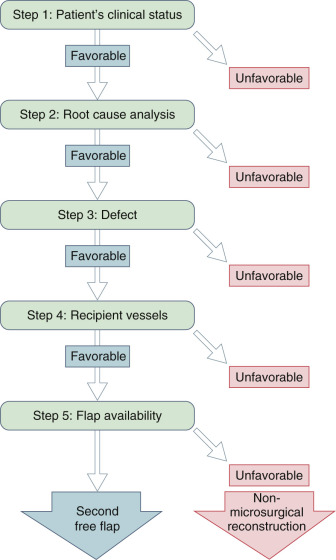
Step One: Clinical Status of the Patient
An honest appraisal of the patient’s medical status is a critical component of the way forward. Typically, the patient has undergone one long primary operation (a resection with a microsurgical reconstruction), an arduous recovery (punctuated with the sentinel event of flap failure), and then a re-exploration for salvage (which has also failed). At this stage, the patient could be quite compromised, and undertaking another long procedure right away may be ill-advised. If the patient is medically unstable, then repeat surgery must be deferred. Even a totally failed flap can serve as a biological dressing for 10 days or more. Most often, this is not necessary, and emergency re-exploration confirms irreversible pedicle thrombosis. At this time, immediate repeat reconstruction is usually performed, especially if loss of the flap results in exposure of vital structures. If, for some reason, repeat major surgery is not advisable, then the necrotic portions of the flap can be debrided and the wound temporarily dressed until a repeat reconstruction can be performed under more suitable circumstances. This course is indicated when the patient’s general medical condition is unfavorable or if preferable for the patient to have an opportunity to participate in a complicated decision-making process. If ultimate failure of all or a portion of the flap would result in an unacceptable recurrent defect, then early debridement of all compromised tissue and repeat reconstruction offers a speedy recovery. If it appears likely that critical portions of the flap have a good chance of survival, then surgery can be delayed to allow maximal tissue salvage and clear delineation of the remaining defect.
Step Two: Root Cause Analysis of Flap Failure
If you don’t ask the right questions, you don’t get the right answers. (EDWARD HODNETT)
When analyzing the causes of free flap failure, a thorough review of microsurgical principles coupled with a candid effort to identify possible breaches in technique is mandatory. This not only helps one to decide whether it is prudent to attempt another free tissue transfer, but it is a discipline that improves the technical abilities of the surgeon.
A root cause analysis (RCA) provides the tools for a systematic approach. Root cause analysis is a structured method used to analyze serious adverse events. Initially developed to analyze industrial accidents, RCA is now widely deployed as an error analysis tool in health care. A central tenet of RCA is to identify underlying problems that increase the likelihood of errors, while avoiding the trap of focusing on mistakes by individuals. RCA thus uses the systematic approach to identify both ACTIVE ERRORS (errors occurring at the point of interface between humans and a complex system) and LATENT ERRORS (the hidden systemic problems that contribute to adverse events). This step is critical to decision-making for the secondary reconstruction.
If failure is attributable to a clearly avoidable cause, then undertaking a second free flap reconstruction is feasible. In sharp contrast, if the cause is an unavoidable problem, a more conservative approach may be the only way the defect can be reconstructed.
In order to help elucidate the issue of avoidable versus unavoidable causes of failure, a brief synopsis of the common causes is presented.
All free flap failures ultimately result from platelet aggregation at the microvascular anastomosis (in patients with normal clotting function). The list of factors that lead to this sentinel event is classified into three main categories ( Box 30.1 ).
Systemic Factors
- •
Low flow states
- •
Drugs
- •
Hypoxia
- •
Comorbid disease
- •
COPD
- •
Hypercoagulable states
- •
Impaired wound healing state
- •
Diabetes
- •
Renal failure
- •
Stroke
- •
Steroid use
- •
Extraluminal Factors
- •
Pressure
- •
Hematoma
- •
Postop positioning
- •
Dressings, tapes, masks
- •
Catheter drains
- •
Edema
- •
Fibrosis
- •
Twist/kink
- •
Tissue tension
Intraluminal Factors
- •
Intimal inversion
- •
Atherosclerosis
- •
Rheologic currents
- •
Embolism
- •
Suture exposure
- •
Radiation
- •
Vascular malformations
- •
Thrombophilia
Systemic Factors
Typically, the systemic factors are directly related to comorbid conditions that the patient suffers and which adversely affect flap survival. Low-flow states , such as cardiac disease, labile hypertension, dehydration, renal failure, and perioperative hypovolemia, vasoactive medications, or recreational drugs that result in vasoconstriction, hypotension, or altered flow to the flap; hypoxia (from COPD), or restrictive pulmonary disease, are possible causes.
Usually they constitute unavoidable causes of flap failure (an exception being perioperative hypovolemia and dehydration, which are correctable) – and steer decision-making away from another free flap reconstruction that could fail again. In most patients with systemic causes of failure, it is safer to choose a non-microsurgical simpler reconstructive strategy and return for a more definitive reconstruction after the factors that led to flap failure have been mitigated. Systemic disorders are associated with poor wound healing or the unacceptable risks of morbidity such as myocardial infarction, stroke, respiratory failure, etc. Flap failure is related to conditions that cannot be immediately reversed such as severe malnutrition or morbid obesity pose a circumstance in which a non-microsurgical alternative may be most prudent. Each patient must be individually assessed for the possible effect of comorbid conditions and the risk and benefits of repeat microsurgery must be thoughtfully weighed. Equally important is to consider the patients’ functional recovery in the setting of comorbid conditions that the patient has inherently.
Extraluminal Factors
Even a technically perfect microanastomosis will thrombose if flow is reduced in the hours and days following surgery. The most common cause of reduced flow is mechanical compression of the vascular pedicle . Largely, these factors arise from the local tissues directly adjacent to the flap, and cause vascular compromise. They typically include external pressure from catheters, tape, dressings, tissue edema, improper positioning, and hematoma formation adjacent to the vascular pedicle. Last but not the least, prolonged surgery may add to the risks of complications.
Intraluminal Factors
Vascular disease involving the donor or recipient vessels may render them prone to thrombosis, despite optimal microsurgical technique and postoperative management. Intima-media thickening, the precursor lesion of atherosclerosis, may begin in adolescence, but definite atherosclerosis tends to appear after the age of 40 in men, and the onset of menopause in women. In the carotid system, a common recipient vessel location for head and neck free flaps, the incidence of asymptomatic atherosclerosis can be as high as 80%, depending on patient age, gender, and associated hypertension, cigarette smoking, and diabetes mellitus. Incidence in the lower extremities is similar. Radiotherapy increases the risk of vascular disease in vessels located in the treatment fields. Patients with vascular malformations can also pose problems due to abnormal local vasculature and clotting abnormalities. Performing microvascular surgery on diseased vessels requires meticulous attention to technical details. When a flap fails despite proper technique and adjuvant systemic anticoagulation, then it is usually prudent to salvage the reconstruction using non-microvascular methods.
Thrombophilia
Hypercoagulable states are associated with a variety of inherited and acquired conditions affecting platelet function or peptides in the clotting cascade. Inherited disorders include conditions such as factor V Leiden, protein C and S deficiencies, heparin-induced thrombocytopenia (HIT; especially when heparin is used indiscriminately either during or after the operation), and antithrombin and prothrombin mutations. Although uncommon, they may account for thrombosis that is difficult to control in surgical patients and lead to poor outcomes. Acquired hypercoagulability is associated with trauma, infection, severe emotional distress, collagen vascular diseases, and cancer. Hormonal contraceptives and replacements promote thrombophilia in women. Cancer, particularly breast and head and neck, is associated with disorders of hemostasis related to abnormal activation of the coagulation pathways, enhanced platelet aggregation and activation, and decreased synthesis of antithrombin III and protein C. Head and neck cancer patients can have both preoperative hypercoagulability and postoperative thrombophilia due to alterations in clotting profiles related to the underlying malignancy as well as major surgery. If significant disorders of hemostasis are suspected or confirmed following a failed free tissue transfer, then repeat microvascular procedure has a high likelihood of a poor outcome and an alternative procedure should be considered.
Step Three: Characterize the Defect
An essential part of planning any reconstructive procedure is to identify the specific anatomic and functional elements involved in the defect and prioritize them for repair. Four critical questions arise:
- 1.
What is the location of and anatomic complexity of the defect?
- 2.
Are vital structures exposed?
- 3.
Is it associated with a functional disability?
- 4.
What is the quality of the tissue bed?
In some sites (e.g., head and neck), functional priorities may take precedence over aesthetic ones. In these situations, early closure is imperative and non-microsurgical salvage may be a quicker and effective solution. In others (e.g., breast reconstruction) aesthetic goals may take precedence. It is never possible to achieve every conceivable goal for a perfect restoration. There are always trade-offs. Thus, the task of the surgeon is to consider which objectives are most important, formulate a menu of surgical options, and choose a method most likely to achieve the essential goals in the simplest fashion with the least risk.
After a failed free flap, the patient again has an unreconstructed or “recurring” defect that requires a diverse armamentarium of reconstructive options. The surgeon must face the same set of decisions regarding treatment but is armed with more information about the patient and perhaps a different set of options from which to select. Decision-making mandates even greater thoughtfulness, and some original goals may have to be abandoned when planning the salvage procedure. A good example is treatment of an irradiated wound. A vascularized tissue transfer is more reliable than a split-thickness skin graft or primary closure in these circumstances. For this reason, a free tissue transfer may be selected for a primary reconstruction. In the event of failure due to irreversible factors, the surgeon might be forced to resort to skin grafting and accept decreased reliability, function, and aesthetic results. An important consideration is the consequence of failure. Failure of a split-thickness skin graft on a wound consisting of muscle and subcutaneous tissue is less consequential than if failure leads to exposed vital structures (e.g., major vascular structures, dura mater, etc.). If the consequences of failure are low risk, then an acceptable trade-off can be to use a less reliable method of reconstruction. If the consequences are unacceptable, then every effort must be made to perform a repeat microsurgical reconstruction under optimized circumstances. It is difficult to generalize about these decisions. Every case must be individually evaluated.
Step Four: Assess the Recipient Vessels
If appropriate donor tissue is available, then recipient vessels are selected. This is the most important decision in a repeat microsurgical operation. Previously used vessels may be considered if flap failure cannot be attributed to some feature associated with them such as inadequate flow or vascular disease. If prior failure was related to small, low-flow recipient vessels, then a repeat free flap should be planned, based on large, high flow vessels. In certain circumstances, vein grafts to high flow vessels (e.g., external carotid artery or common femoral artery) can be more reliable than a direct pedicle anastomosis to small recipient vessels. They can also be used to reach locations away from vessels affected by vascular disease caused by atherosclerosis or radiation. The flap pedicle itself can be used as recipient vessels if flap failure involves only a portion of the flap (e.g., the skin paddle of a composite bone flap), or if it is related to factors intrinsic to the flap design and not to the microanastomosis. If this option is chosen, then it is best to delay surgery until after the endothelium has healed (i.e., 7 days), so there is less chance for thrombosis when flow is stopped during repeat surgery.
If suitable donor tissue and recipient vessels are available and a repeat microvascular reconstruction is planned, then careful attention must be given to avoiding all ancillary possible factors that might have contributed to the previous failure. Anticoagulation is not necessary unless indicated based on previous influences based on diseased vessels or coagulopathies.
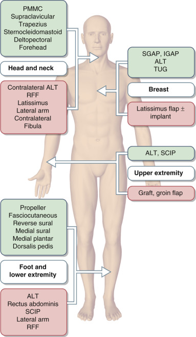
Step Five: Examine Reconstructive Choices
More complicated defects are associated with longer lists of possibilities. The critical question always is will the functional and cosmetic goals be met following the second surgery? Alternative donor sites can be selected to contain similar tissues as the original donor area but with adjustments made based on possible factors that might have contributed to failure associated with previous donor site selection ( Fig. 30.2 ). Donor areas supplied by short, small vessels (e.g., sophisticated perforator flaps) may be altered to include more tissue and longer, large-caliber vessels. If this results in a reconstruction with excessive soft tissue bulk, then plans can be made for subsequent revisions. Composite tissue flaps (e.g., fibula osteocutaneous free flaps) may be simplified to flaps containing only soft tissues (e.g., rectus abdominis musculocutaneous flaps), ALT for breast reconstruction. Propeller flaps based on pivoted perforator vessels are elegant options for salvage. In head and neck reconstruction, the trade-off in this decision may be a less functional reconstruction but greater chance of achieving a healed wound with vital structures protected, oral competence, and prevention of an orocutaneous fistula. In some cases, a portion of the flap survives (e.g., necrosis of only the skin component of a fibula osteocutaneous free flap) and a repeat reconstruction has less extensive requirements than the original. Often, it is appropriate to consider contralateral harvest of the same type of flap used in the original surgery.