(1)
Yotsuya Medical Cube, Chiyoda-ku, Tokyo, Japan
Basic Principles
The tissue required for reconstruction of the calcaneal region is thick and must be able to withstand weight applied. It is preferable to have sensory function, but it is not necessary to pro-actively reconstruct sensory function, so tissue that can obtain protective sensation easily is selected.
For this reason the medial plantar flap is the first choice. Only if for some reason it is not possible to obtain the medial plantar flap from the affected foot or from the healthy foot either, another flap such as an abdominal perforator flap should be examined.
Whichever flap is selected, guidance must be given for long term use of an arch support splint.
Selectable Flaps and Surgical Procedures
Medial plantar flap (Normal/free)Medial pedis flapFree scapular flapFree abdominal perforator flapLateral calcaneal flapReverse peroneal flapAnterolateral thigh flap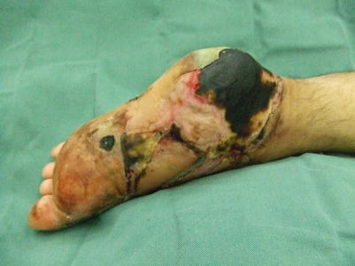
18.1 Medial Plantar Flap (Level of Difficulty: 3)
Information
Vascular pedicle Superficial branch of medial plantar artery
Size From the upper plantar arch area to the periphery of the scaphoid tubercle
Advantage Suitable for reconstruction of plantar weight bearing area. Possible to include sensory function
Caution If the stitching is located on the weight bearing section it can produce new scar contracture. Therefore sufficient examination is required prior to surgery to ensure that the flap harvest area and stitching post-surgery does not occur in the load bearing area.
For reconstruction exceeding the size of the upper arch, it is better to examine the possibility of using other free flaps. Also, guidance should be given to the patient for the use of an arch support splint for several months after surgery. This is not suited to cases where patients are required to walk on gravel roads or wear thin rubber soled shoes as part of their work.

18.1.1 Operation Procedures

Fig. 18.1
Procedure 1: Large skin defect present on lateral plantar weight bearing area. A triangular flap with the same width as the skin defect area is designed from the medial plantar to the upper arch

Fig. 18.2
Procedure 2: Scar from around the skin fistula is completely removed

Fig. 18.3


Procedure 3: An incision is made around the margin of the skin flap, and the skin flap is detached while retaining the toe nerve beneath the plantar fascia. The tarsal tunnel is opened from the proximal end and the vascular pedicle is detached while retaining the calcaneal branch/lateral plantar artery at the distal end of the posterior tibial artery. The abductor hallucis muscle is retained and the vascular pedicle is detached from beneath it. Sensory nerves are included in the skin flap if they can be retained
Related posts:


Stay updated, free articles. Join our Telegram channel
Full access? Get Clinical Tree
