Raynaud disease and phenomenon


Raynaud disease and phenomenon


Sameh S. Zaghloul, Najat A.Y. Marraiki and Mark J.D. Goodfield


Evidence Levels:  A Double-blind study  B Clinical trial ≥ 20 subjects  C Clinical trial < 20 subjects  D Series ≥ 5 subjects  E Anecdotal case reports


image


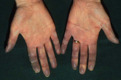
Raynaud phenomenon (RP) is characterized by intermittent peripheral vasoconstriction leading to pallor, cyanosis, and reactive vasodilatation of the arterioles of the fingers and toes. It is caused by vasospasm in response to cold, emotion, hormones, and certain vasospastic drugs. Primary Raynaud disease is a milder, idiopathic form, whereas secondary RP coexists with autoimmune connective tissue disorders such as systemic lupus erythematosus and systemic sclerosis, or other conditions that reduce blood flow, such as localized structural abnormalities.


Despite the increases in our understanding of disease mechanisms involved in Raynaud disease, the precise pathogenesis is not fully understood. The pathogenesis and pathophysiology vary between the primary (idiopathic) and the secondary forms.


Management strategy


Raynaud disease is often mild and may not require treatment; the use of warming devices such as hand or toe warmers is beneficial; however, with secondary Raynaud disease there is not only vasospasm but often fixed blood vessel damage, so the ischemia can be more severe. Complications can include digital ulcers and could, rarely, lead to amputation. Treatment is often non-pharmacological including avoiding cold and smoking cessation. Calcium channel antagonists, such as nifedipine (10–60 mg daily), are often considered when treatment is needed; however, the adverse effects of these drugs can include hypotension, vasodilatation, peripheral edema, and headaches. Other treatments that have been studied in randomized, controlled trials include classes of drugs such as angiotensin II inhibitors, selective serotonin reuptake inhibitors, phosphodiesterase-5 inhibitors (e.g., sildenafil, 25–50 mg up to four times a day), and nitrates (topical or oral). For more serious Raynaud disease or its complications, prostacyclin agonists may be used. This may be particularly useful for RP associated with connective tissue disease. There are also studies demonstrating that endothelin receptor blockade with bosentan (62.5 mg bid) can reduce the number of new digital ulcers in scleroderma patients. However, it does not affect the healing period, and has no effect on the number and severity of attacks of RP in those without ulcers. Natural remedies such as Ginkgo biloba have also been advocated. Avoidance of triggers such as cold (especially sudden drops in temperature) and vibration (in cases where vibration is the precipitant) should be stressed. Drugs that may exacerbate the condition include β-blockers, bleomycin, caffeine, cisplatin, ergot preparations, interferon, methylsergide, nicotine, oral contraceptives, reboxetine, tegaserod, and vinblastine and should be avoided.


Specific investigations


Careful history taking and clinical examination followed by investigation to detect potential underlying disease are essential: capillaroscopy and specific autoantibody tests are the most productive in aiding diagnosis.


Assessment of nailfold capillaroscopy by × 30 digital epiluminescence (dermoscopy) in patients with Raynaud phenomenon.

Beltran E, Toll A, Pros A, Carbonell J, Pujol RM. Br J Dermatol 2007; 156: 892–8.


The sclerodermic pattern showed a sensitivity of 76.9% and a specificity of 90.9% in SS. A typical capillaroscopic pattern of SS was observed in 73% of cases of limited SS and in 82% of cases of diffuse SS. Patients with Sjögren syndrome and dermatopolymyositis-SS showed a non-specific capillaroscopic pattern. All patients with primary RP presented a normal capillaroscopic pattern. A normal capillaroscopic pattern was also observed in 11 of 12 patients with pre-SS. Digital epiluminescence seems to be a useful and reliable technique in the evaluation of capillary nailfold morphological changes. This technical variation allows the identification of specific capillaroscopic patterns associated with connective tissue diseases. It also permits us to differentiate primary RP from secondary RP. The results obtained with this technique are similar to those previously reported using standard capillary microscopy, but this is much easier.


First-line therapies


Stay updated, free articles. Join our Telegram channel

Aug 7, 2016 | Posted by in Dermatology | Comments Off on Raynaud disease and phenomenon

Full access? Get Clinical Tree

Get Clinical Tree app for offline access