Racial Considerations: Skin of Color: Introduction
|
Racial and Ethnic Influences on Skin Disease and Therapy
In the United States and worldwide, myriad cutaneous phenotypes characterize mankind. Most striking is the range of skin and hair color (Fig. 9-1). The Census Bureau estimates that half of the US population will be of non-European descent by the year 2050.1 There are currently more than 95 million persons in the United States2 and billions of individuals worldwide categorized as having “skin of color.” There has been increasing awareness of racial and ethnic influences (see Table 9-1) on skin biology and on diagnosis and treatment of skin disease.

Term | Derivation | Current Usage or Implication |
|---|---|---|
Race | < Latin, root | A population distinguished as a more or less distinct group by genetically transmitted physical characteristics; originally geographically segregated Groups increasingly blurred by interracial offspring Determined at conception, immutable May influence disease predilection/susceptibility |
Ethnicity | < Greek, race or distinct population with its own customs | A social construct in which a group of individuals shares a language, cultural practices, or customs Self-assigned and somewhat mutable Often but not always associated with race May influence treatment choices and exposure to disease-promoting or disease-preventing factors |
The literature regarding “skin of color” primarily focuses on promoting awareness of normal and abnormal skin conditions in a patient regardless of skin phenotype. It seeks to identify risks and benefits of treatments in diverse skin types, to develop effective treatments for common dermatoses in skin of color, recognizing the importance of individualized therapy, and to avoid stereotyping and generalization.
Defining Skin of Color
In defining skin of color, it is important to consider the reasons for doing so. Many have questioned the common propensity of medical practitioners to state a patient’s race among the first few words, as a primary identifier. The skin type, color, or ethnic background of most patients may be better suited to the physical examination or to relevant points in the history. The International Committee of Medical Journal Editors’ Uniform Requirements for Manuscripts Submitted to Biomedical Journals recommends that authors using variables such as race or ethnicity should “define how they measured the variables and justify their relevance.”6 The Journal of the American Academy of Dermatology similarly suggests that authors inclined to submit racial, ethnic, or skin color descriptors in manuscripts ask themselves a series of questions regarding whether such identification is important to the understanding or pedagogical value of the manuscript, whether the patient would self-identify in the same way and how this is known, whether the descriptor used could be open to racist interpretation, and what the evidence is that the descriptor plays a role in the entities described.7
Most would argue that “nonwhite” skin is “skin of color.” However, there is a diverse array of phenotypes within the nonwhite and white spectra, and two categories are inadequate to describe them. The most commonly used classification system in dermatologic practice is the Fitzpatrick phototype,8,9 designed to provide an estimate of skin cancer and photoaging risk, in which individuals are assigned a number based on their ability to burn and tan after a single, rather modest sun exposure. The original Fitzpatrick system consisted of types I–IV; types V and VI were soon added with the goal of representing individuals of Asian/Indian and African/Aboriginal origin, respectively. Skin of color is now commonly regarded as encompassing types IV through VI,10 but many patients and physicians would argue that the spectrum is broader. For example, Japanese women not infrequently describe themselves as having type II skin. In any case, for the purpose of studying nonwhite skin, the Fitzpatrick system has several limitations. Although intended to utilize patient-reported characteristics, in practice a patient is commonly “assigned” to a type without benefit of a history. Also, the Fitzpatrick type has been shown in studies not to correspond well to constitutive skin color and to correlate poorly with minimal erythema dose (MED) values.11 Interestingly, the physician-assigned skin phototype has been shown to correlate moderately with race, and race assignment does not correlate well with objective measures of pigmentation or self-reported skin phototype, suggesting that assignment of Fitzpatrick types is often based on perceived race rather than either objective skin color or response to ultraviolet (UV) light.12
Other scales have been developed with a goal of more accurately defining skin pigmentation and corresponding to defined clinical features. It has been suggested that for skin of color a scale based on propensity to postinflammatory hyperpigmentation, for example, might be more useful than one based on burning and tanning responses to UV exposure.10 A variety of visual scales seek to convey skin color more accurately and precisely and to measure changes over time. These include the physician’s global assessment, melasma area and severity index (MASI), visual hyperpigmentation scale,13 and skin tone color scale.14 Skin photometers employing UV light, polarized light, reflectance spectroscopy, tristimulus colorimetry, and spectrophotometry are also available and can provide an objective skin color score.13 However, it is possible that, parallel to the Fitzpatrick phototype classification for white skin, a classification system based on patient perceptions of desirable or adverse responses to environmental challenges would be most useful in managing dermatologic patients with skin of color. For example, asking the patient about the frequency, severity, and duration of postinflammatory hyperpigmentation in the past may provide critical guidance when considering a skin care or treatment regimen.
Structure and Function in Skin of Color
The perceived skin color is the result of the spectrum of incident light and its absorption and reflection by chromophores in the skin. It is largely determined by melanin amount, type (ratio of black/brown eumelanin to red/yellow pheomelanin), and intracellular distribution and location within the layers of the skin. The number of melanocytes in a given area of skin is similar among all individuals. Vascular pigments oxyhemoglobin and deoxyhemoglobin also play a role in observed color along with capillary blood flow, dietary pigments carotene and lycopene, collagen, the spectrum of ambient light, reflection, refraction, and absorption of light by the skin, and transparency of the stratum corneum and epidermis.15 Endocrine, inflammatory, neural, and pharmacologic factors also influence skin color.
Melanosome size and distribution vary in skin of differing colors (Fig. 9-2). In individuals of African descent, the melanosomes are typically large and dispersed singly in the keratinocytes. In white individuals, melanosomes are smaller and grouped within a membrane. In Asian skin, a combination of individual and clustered melanosomes is found.16 Melanin may also be degraded more slowly in skin of color.17 Increasing melanin provides higher natural photoprotection, due to a greater absorption of UV photons, but also typically increases risk of pigmentary disorders including hypopigmentation or hyperpigmentation as a result of physiologic responses to trauma or inflammation.18 For further discussion, see Chapter 72.
Figure 9-2
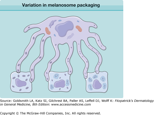
Variation in melanosome packaging within the keratinocyte of skin of differing colors. White skin (right) typically shows spherical melanosomes clustered within a membrane, black skin has larger elliptical melanosomes dispersed singly, and Asian or golden brown skin typically has a combination of the two (middle). (Used with permission from Jag Bhawan, MD.)
Structural or functional differences in white versus nonwhite human skin beyond those related to photoprotection19 and pigmentary alterations are not known. Conflicting data have been presented on variations in lipid content, sebum content, and the number of stratum corneum layers and compaction.20 Histologic sections of normal skin generally appear identical aside from the difference in melanin. Whether there are genetically determined functional differences of the skin associated with skin color is the subject of study and debate.15
Genetics of Pigmentation
There is a polygenic basis for human pigmentation. Genes already implicated in pigmentary variation of human skin, hair, and iris include TYR, TYRP1, OCA2/HERC2, SLC45A2 (OCA4/MATP), SLC24A5, SLC24A4, MC1R, ASIP, KITLG, IRF4, and TPCN221,22 (Table 9-2).
Gene | Gene Product | Recognized Function | Pigmentary Variation Effect |
|---|---|---|---|
TYR | Tyrosinase | Rate-limiting enzyme in melanin biosynthesis | Europeans and South Asians: lighter/darker skin within group; little variation among races |
TYRP1 (TRP1) | 5,6-Dihydroxyindole-2-carboxylic acid oxidase or tyrosinase related protein 1 | Enzyme in melanin biosynthesis | Europeans: lighter/darker skin and eye color |
OCA2 (HERC2) | P protein | l-Tyrosine transmembrane transporter | Blond/brown hair, blue/nonblue eyes; East Asians, Africans: lighter/darker skin Loss of function causes oculocutaneous albinism type 2 |
SLC45A2 (MATP) | Membrane-associated transporter protein | Transmembrane transport | Black/nonblack hair; dark/light skin; dark/light eyes |
SLC 24A4 | Sodium/potassium/calcium exchanger 4 | Transmembrane potassium-dependent sodium/calcium exchanger | Blond/brown hair; blue/green eyes |
SLC24A5 | Sodium/potassium/calcium exchanger 5 | Transmembrane potassium-dependent sodium/calcium exchanger | Africans and Asians: ancestral form (111Ala), darker skin; Europeans: variant (111Thr), lighter skin |
MC1R | Melanocortin 1 receptor | Receptor for melanocyte-stimulating hormone (MSH) and ASIP | Red hair–fair skin: R151C, R160W, D294H mutations; loss of function increases pheomelanin |
ASIP | Agouti-signaling protein | Ligand for MC1R; antagonist for MSH that increases pheomelanin | Inactivating mutations associated with dark hair/eyes |
KITLG | KIT ligand (steel factor, stem cell factor, mast cell growth factor) | Ligand of KIT tyrosine–kinase receptor (cell-migration effects) | Blond/brown hair; lighter/darker skin |
IRF4 | Interferon regulatory factor 4 | Regulator of RNA polymerase II transcription factor activity | Light/dark hair |
TPCN2 | Two-pore calcium channel 2 | Lysosomal membrane calcium channel | Blond/brown hair |
Examination of mitochondrial DNA and Y chromosome analysis suggests that all humans descended from three African females and three African males.22,23 It is believed that some of these descendants stayed in Africa, while others migrated to Europe and Asia and from Asia across the polar land bridge to the Americas. West Africans with the ancestral variant of the SLC24A5 gene expressing alanine at amino acid 111 have dark brown skin. East Asians also have this form of SLC24A5.24 A variant of the SLC24A5 gene with threonine at amino acid 111 is nearly constant in those of European ancestry, determining lighter skin types, and is believed to have resulted from natural selection.24 SLC24A5 encodes a potassium-dependent sodium/calcium exchanger that is hypothesized to play a role in melanosome morphogenesis and melanogenesis through changes in intraorganellar calcium concentration and pH. The SLC24A5 variation is believed to explain 25–38% of the melanin difference between Africans and Europeans.24 Polymorphisms of SLC45A2, OCA2, and KITLG have also been shown to contribute to pigmentation differences between European and African populations.25
Within European-derived populations, TYR, OCA2, MC1R, ASIP, and IRF4 polymorphisms account for much of the observed pigmentary variation.25 The best studied concerns the highly polymorphic MC1R, a melanocyte surface receptor that when activated by its ligand aMSH increases intracellular cAMP levels, induces the microphthalmia transcription factor MiTF and ultimately increases black/brown eumelanin synthesis. A number of loss-of-function variant alleles of MC1R have been identified and shown to result in a fair-skinned red-hair phenotype.26 Other MC1R variant alleles result in blond or light brown hair and fair skin.26 Conversely, dark brown or black hair and dark skin are observed in individuals expressing the wild type MC1R. Risk of skin cancer, including melanoma, is also affected by the inherited MC1R alleles and is generally correlated with fair skin color.26 However, some variant alleles appear to confer an increased risk independent of their effect on pigmentation.26
No genetic basis for the different pigmentary phenotypes of Africans compared with Asians has yet been identified. Both have the ancestral form of SLC24A5 and MC1R, suggesting that evolution to lighter skin colors may have occurred independently in Europe and in Asia. The genes regulating pigmentation differences among Asians are not adequately characterized, although OCA2 variant alleles have been associated recently with some East Asian skin pigmentation variations. South Asian skin pigmentation shows considerable variation; variants of SLC24A5, SLC45A2, and TYR have been associated with these pigmentary differences.27
The “Hispanic” skin color group is least well defined. It is genotypically and phenotypically variable, representing mixtures of European (largely Spanish), African, and Central and South American Native origins. The Hispanic phenotype often differs on average in different geographic areas. Perhaps because of these challenges, genetic determinants of Hispanic skin color remain virtually unstudied.
Variation in Hair Characteristics
Human hair is typically categorized into three major distinct groups: (1) Asian, (2) Caucasian, and (3) African.28 However, the world’s population encompasses people of multiple and mixed backgrounds, resulting in the existence of multiple hair types.29 All hair exhibits common characteristics of morphology, chemical makeup, and molecular structure. There are nevertheless differences in hair morphology and physical properties that contribute to the unique features of the hair fiber, response to hair treatments, and development of disease processes in different groups.30 Understanding the biology and the differences in physical properties of various hair types can assist clinicians in managing hair and scalp problems. A variety of terms are used in the dermatologic literature to describe different hair types based upon an author’s personal preference, country of origin, or current trends. To date, most studies have focused on African hair, which presents the greatest array of clinical disorders.
African or black hair is known to be more affected by breakage, with easily observed fragility in vivo.31 There are no known chemical differences in black versus Caucasian or Asian hair to explain this observed fragility. The biochemical composition of hair in people from different geographic regions and racial groups has been shown to be virtually identical in terms of keratin and amino acid content,32,33 despite significant differences in tensile strength, combability, and moisture content. In contrast, numerous studies have described the physical differences in hair from people of different races.34–37 As well, African-Americans have a significantly lower hair density (number of follicles per unit area) than whites (22.4 vs. 35.5 follicles); and a study examining Asian scalp biopsies found lower hair density than in Caucasians.38 Such differences must be taken into account in the interpretation of scalp biopsy specimens.39
Initially, the cross-sectional shape of the hair shaft was thought to determine the macroscopic appearance of hair and to distinguish people of different genetic backgrounds.40 The round cross-section of Asian hair was thought to result in straight hair, the elliptical or flattened cross-section of African hair to result in curly hair and an intermediate to round and elliptical cross-section results in wavy to straight European hair (Table 9-3). However, three-dimensional computer-aided reconstructions of scalp biopsy cross-sections suggest that the shape of the hair follicles (helical or curved in Africans vs. straight and perpendicular to the skin surface in Asians) also play an important role. The finding that Caucasians can have hair follicles that are elliptical in cross-section and yet have straight hair shafts further implies that the three-dimensional structure of the hair follicle is responsible for the shape of the hair shaft. In vitro experiments comparing the growth of curly and straight hair found that follicles producing curled hairs, when dissected out of the scalp and placed in culture, continue to grow curled hair shafts.41 This suggests that the shape of the hair may be genetically programed by the bulb, with or without the usual dermal environment.42
Racial Group | African | Caucasian | Asian |
|---|---|---|---|
Cross-sectional shape | Elliptical | Ovoid | Round |
Hair follicle shape | Curved | Variable | Straight |
Tensile strength | Low | High | High |
Work of combing | High | Low | Low |
Moisture content | Low | High | High |
Average number of cuticle layers | Highly variable (6–8 along the major axis, 1–2 along the minor axis) | Constant (4–5) | Constant (6–7) |
Recent studies have identified Asian-specific alleles of the EDAR and FGFR2 genes that are associated with thick, straight hair.43,44 Likewise, a recent genome-wide association scan for hair morphology (straight, wavy, curly) in Australians of European descent suggested that polymorphisms of the trichohyalin gene (TCHH), which is expressed in the developing inner root sheath of the hair follicle, contributes to the variance in hair morphology.45 A quantitative study examining hair formation in seven populations demonstrated African hair to have the highest amount of curvature and kinking and a periodic widening and narrowing of the cuticle layer. The cuticle layer of curly hair averages 6–8 layers thick at the end of the major axis but only 1–2 layers at the ends of the minor axis, a weak point susceptible to hair damage from mechanical and chemical forces.46 A study of cuticle differences between Asian (Koreans specifically) and Caucasian hair revealed a 40% larger mean hair diameter, an increased number of cuticle layers, and a thicker cuticle layer in Asian compared to Caucasian hair (Table 9-3).47 Cuticle thickness may play a role in determining the different characteristics of hair, such as chemical reactivity to hair coloring or perms, resistance to UV radiation, and mechanical resilience.
Differences have also been observed in the rate of hair growth. Although hair from individuals of European descent has been observed to grow an average of 1 cm a month,48 African hair grows an average of 0.77 cm a month.49–51 In a comparative light and scanning electron-microscopic study of African, European, and Asian hair, African subjects who did not have a hair cut for over 1 year had hair lengths significantly shorter than would be expected at a growth rate of 1 cm per month. Possible contributors to the differences in hair length, other than a slower growth rate, could include a significantly shorter anagen phase of the hair cycle or perhaps repeated breakage of African hair. In the same comparative study of African, European, and Asian hair, the African hair appeared as a tightly coiled spring-like structure. Compared with shafts from other ethnic groups, many shafts contained trichonodosis or knots (10–16% vs. 0.15%) and other shafts appeared broken (eFig. 9-2.1). The study found a lower incidence (<40%) of hairs with attached roots in the African hair samples compared with more than 75% and approximately 90%, respectively, for the Caucasian and Asian samples; and a greater incidence of tips with frayed or serrated appearance when compared to Asian and Caucasian hair that had cut tips. These data suggest that most of the African hair collected from combing in this study was broken and not shed.51 This raises the question of whether African hair is breaking off during grooming because of increased fragility.
eFigure 9-2.1
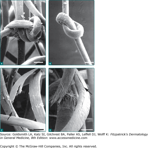
A. Detail of knot in the African hair. Note complex nature of the knot with damage to the cuticle exposing the cortical fibers. B. Detail of the only knot observed in the Caucasian hair, which appears to be looser with no damage to the cuticular layer. C and D. Details from African hair mat shows the longitudinal fissures of the shafts (arrows) plus examples of splitting (C) and breaking (D) of the hair shaft. (A–D, Scale bar = 0.1 mm). (Reproduced with permission from Khumalo NP et al: What is normal black African hair? A light and scanning electron-microscopic study. J Am Acad Dermatol 43(5 Pt 1):814-820, 2000.)
Hair fragility is measured using tests of tensile strength, and it has been suggested that the force needed to break African hair fibers is less than that for other population groups. In a review of tensile strength tests obtained from four different private laboratories, two showed no difference between African hair and that of Caucasian and Asian hair and the other two found African hair to be weaker. Others found a lower tensile strength of both wet and dry curly African hair compared to wet and dry Caucasian hair.
The strength of hair has been shown to be dependent upon the integrity of its sulfur-rich proteins and disulfide bonds. In a study of trichothiodystrophy (TTD), a condition characterized by reduced sulfur-rich proteins and increased hair fragility, control hairs from African, Asian, and Caucasian subjects had statistically comparable sulfur staining using transmission electron microscopy and specific sulfur stains, while hair of patients with TTD was distinct.52 Examination of cultured curly hairs in vitro showed a variable thickness of the outer root sheath, which was thicker on the concave side of the follicle, indicating some alteration in the differentiation process of hair compartments. It is unclear whether the lack of symmetry of the African hair bulb increases the tendency to mechanical damage, but it is likely that the shape of African hair makes it susceptible to physical damage as a result of certain grooming practices. In addition, intraracial variation in the degree of curl may influence mechanical properties. In a comparative study of African-American hair with different degrees of curl, from a loose to a tight curl pattern, mechanical fragility of hair increased with a tighter curl pattern.53 Certain ethnic hair care practices, such as repeatedly subjecting hair to extremely high temperatures or processing with chemical straighteners, may further damage African hair.
In a study examining the amount of work required (measured in joules) to pass a comb through locks of hair, it was found that the work of combing wet African-American hair is almost five times that of combing wet straight hair. For dry hair, the work is 50 times greater. The teeth of a comb and method of combing can influence the extent of resulting damage. Broken hairs from combing are more numerous and of shorter length in curly African-American hair compared to straight Caucasian and Asian hair. Knots or trichonodes that are commonly seen in tightly curled hair are sites especially susceptible to damage by comb teeth.
African-American hair demonstrated a slightly lower water content than Caucasian hair.37 The spiraling of the hair shaft may be another reason for increased hair dryness in African-American hair, as sebum from sebaceous glands cannot effectively navigate the twist and turns of the hair shaft, leading to a drier, more brittle hair. The relative “dryness” of African-American hair is worsened by the cumulative effect of environmental forces. Features of such weathering include a damaged cuticle, longitudinal fissures known as “split ends,” and transverse fissures resembling the nodes of trichorrhexis nodosa.
When combing untreated curly hair, a highly negative electrostatic charge develops, in contrast to the low positive electrostatic charge for untreated straight hair. The highly negative charge on African-American hair may be the result of decreased moisture content and increased pulling force from combing. Also, the higher electrostatic charges in African-American hair can produce “flyaway” hair and can lead to difficulty in styling.