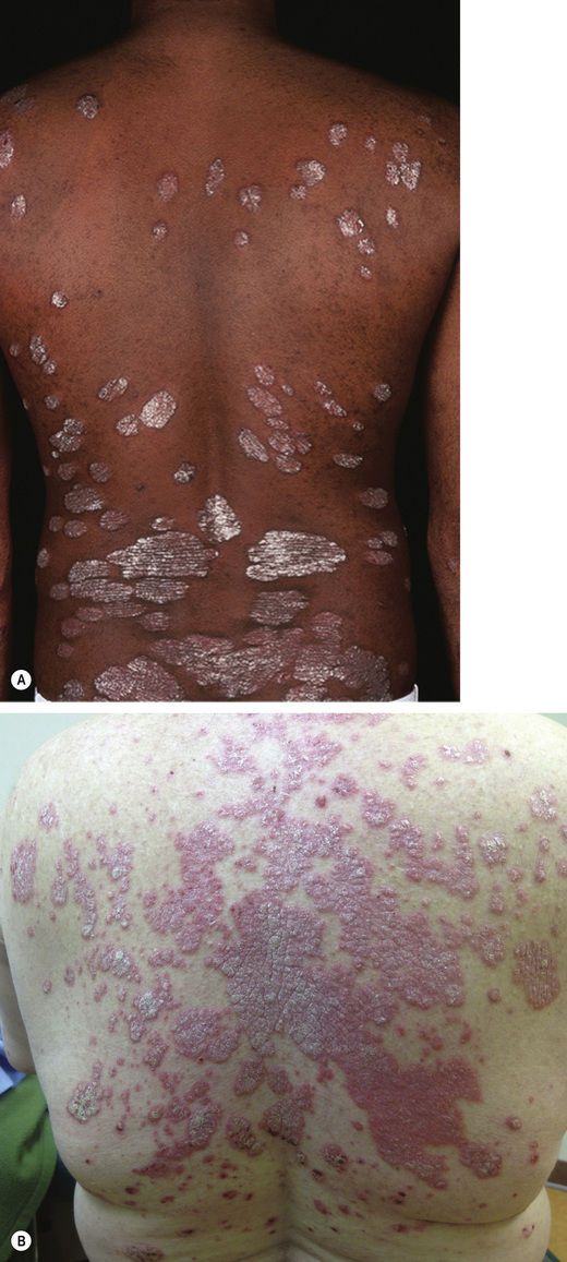
Fig. 4.1Psoriasis, plaque type, most common distribution.
Fig. 4.2Psoriasis, plaque type.A, Courtesy, Peter C M van de Kerkhof, MD. From Bolognia JB, Jorizzo JL, Rapini RP. Dermatology, 2e. London: Saunders, 2008, with permission.
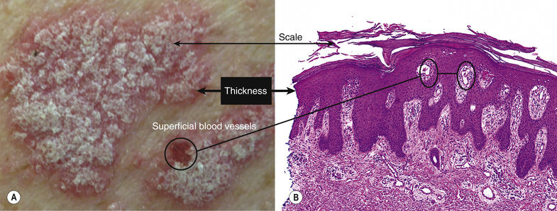
Fig. 4.3Psoriasis.A, From Bolognia JL, Jorizzo JL, Schaffer JV. Dermatology, 3e. London: Saunders, 2012, with permission.
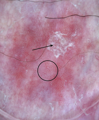
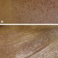
Well-developed scale is silvery (arrow) (Fig. 4.4)
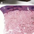
Fig. 4.4Psoriasis (dermoscopy). Silvery scale (arrow) and prominent regular dotted vessels (circle).Courtesy, Giuseppe Argenziano, MD, and Iris Zalaudek, MD. From Bolognia JL, Jorizzo JL, Schaffer JV. Dermatology, 3e. London: Saunders, 2012, with permission.
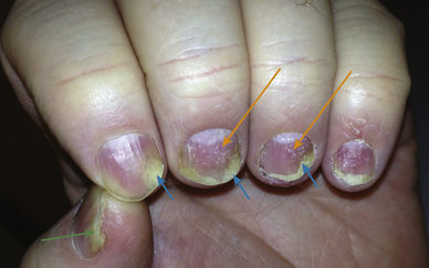
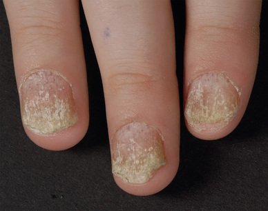
Fig. 4.7Psoriatic nails.Courtesy, Peter CM van de Kerkhof, MD, and Frank O Nestlé, MD. From Bolognia JL, Jorizzo JL, Schaffer JV. Dermatology, 3e. London: Saunders, 2012, with permission.
Psoriasis Variants
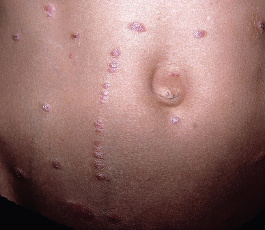
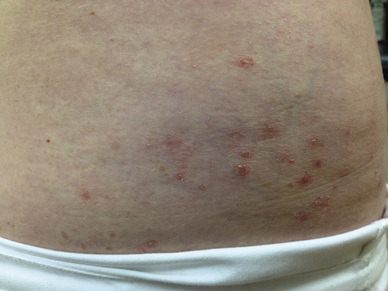
Guttate (see Chapter 5) – small lesions with characteristic scale, generally <1 cm in size (Fig. 4.8)
Fig. 4.8Guttate psoriasis.
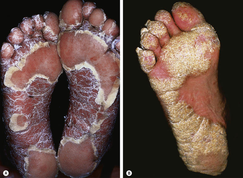
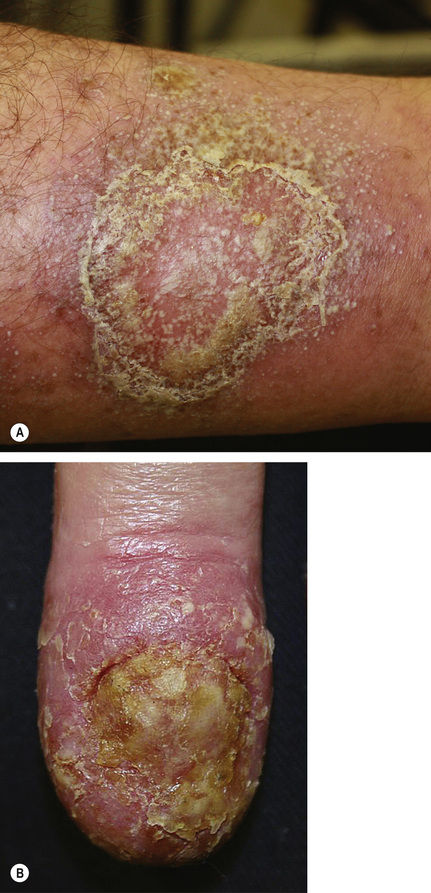
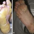
Palmoplantar (see Chapter 2) – lesions with typical scale; underlying skin in these sites may not be erythematous (Fig. 4.9)
Fig. 4.9Plantar psoriasis.A, Courtesy, Peter CM van de Kerkhof, MD. B, Courtesy, Yale Dermatology Residents’ Slide Collection. From Bolognia JL, Jorizzo JL, Schaffer JV. Dermatology, 3e. London: Saunders, 2012, with permission.
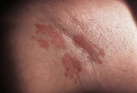
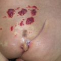
Inverse (see Chapter 2) – minimal scale over thin, pink plaques (Fig. 4.10)
Fig. 4.10Inverse psoriasis.Courtesy, Ronald P Rapini, MD. From Bolognia JL, Jorizzo JL, Schaffer JV. Dermatology, 3e. London: Saunders, 2012, with permission.
Pustular (see Chapter 7) – erythema and pustules; pustules may form “lakes of pus” (Fig. 4.11)
Fig. 4.11Pustular psoriasis. On the finger, this is termed acrodermatitis continua of Hallopeau.Courtesy, Yale Dermatology Residents’ Slide Collection. From Bolognia JL, Schaffer JV, Duncan KO, Ko CJ. Dermatology Essentials, 1e. Philadelphia: Saunders, 2014, with permission.