Side effect
Medical treatment
Procedural treatment
Acne/acneiform eruptions
Topical anti-acne treatment as per severity of acne—benzoyl peroxide, adapalene
Comedone extraction
Chemical peels
Intense pulsed light
Telangiectasia and rosacea
Topical brimonidine
Systemic doxycycline
Intense pulsed light
Pulsed dye laser
KTP laser
Nd:YAG laser
Hypertrichosis/hirsutism
Eflornithine
Long-pulsed Nd:YAG laser
Diode laser
Intense pulsed light
Atrophy/striae
Tretinoin, glycolic acid
Chemical peels, pulsed dye laser, fractional CO2 laser, intense pulsed light, excimer lamp/laser
Subcutaneous atrophy
Topical retinoid. Self-resolving in the early stages may become irreversible in later stages
Hyaluronic acid fillers or autologous fat transfer in irreversible cases at cosmetically important sites
Hypopigmentation
Topical tacrolimus
Excimer lamp/laser
30.2.1 Procedural Techniques for Steroid Abuse
30.2.1.1 Acne and Acneiform Eruptions
Topical steroid abuse in patients with pre-existing acne is common as it leads to a quick reduction in erythema and edema giving an appearance of instant improvement. With prolonged use, patients present with monomorphic erythematous papular and pustular lesions. However the presence of numerous, multiple open and closed comedones, giant comedones, and cysts is not uncommon (Fig. 30.1a, b). In such patients comedone extraction with a comedone extractor leads to faster improvement. In the case of multiple closed comedones, light electrodessication or piercing with a sterile disposable no. 26 G needle, under topical anesthesia, followed by comedone extraction leads to gratifying improvement over a short period. Multiple sessions may be required for complete clearance. Topical and systemic anti-acne medication as appropriate for the severity of acne should be continued. Topical retinoids should be used judiciously as they can cause further irritation to an inflamed erythematous face, due to the steroid withdrawal.
Fig. 30.1
Topical steroid induced acne. (a) Multiple closely packed visible and submarine comedones. (b) erythematous papules and pustules
Chemical peel with a salicylic-mandelic acid combination in a gel base is another useful procedure in resistant cases. The advantage is that salicylic acid leads to reduction in erythema due to its anti-inflammatory effect, and mandelic acid has an antibacterial and skin-lightening effect (Fig. 30.2a, b). The patient should be primed well with a sunscreen before performing peels as photosensitivity is common in patients with steroid abuse. Gel base peels are better tolerated as compared to alcoholic solutions in sensitive skins. Interval between peeling sessions should be longer at 3–4-week intervals to allow for complete healing of the skin between the sessions.
Fig. 30.2
Improvement of acne with comedone extraction followed by combination salicylic -mandelic acid peels
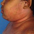
30.2.1.2 Telangiectasia and Steroid-Induced Rosacea
Steroid-induced telangiectasia is a common manifestation of steroid abuse and can be picked up very early with dermoscopy (Fig. 30.3a, b). It is caused by stimulation of release of nitric oxide by the steroid from the endothelial cells of the dermal vessels, leading to abnormal dilatation of capillaries [4]. It can present as redness and close examination on stretching the skin will reveal the dilated capillaries. Treatment is difficult and depends on the size of the vessel. The older procedures used were cauterization of individual vessels with electrosurgery. This was tedious and painful and sometimes led to scarring. The advent of next-generation intense pulsed light (IPL) systems and pulsed dye lasers (PDL) has given rise to better treatment options [5]. Though these lasers do not penetrate deeply, they are effective since most of the vessels are superficial and small (0.5–1 mm) in diameter. Wavelengths used are between 525 and 595 nm. Purpura can occur due to rupture of small vessels during treatment. This can be a problem but usually clears in 1–2 weeks. Using large spot sizes and effective cooling can make side effects less frequent. A typical treatment would include the use of an 8–10 mm spot size, fluence of 6.5–8.5 J/cm2, and 6 ms pulse width. The number of treatments required can vary from one to four done at 4–6-week intervals. Prominent and slightly larger vessels can be treated individually with a 1 mm spot size and short pulses, moving slowly along the vessel. The diffuse redness of rosacea is better treated with the 595 nm pulsed dye laser. Though brimonidine tartrate gel helps in reducing redness, the effect is temporary and only lasts for 12 hours. The other lasers that can be used are the 532 nm KTP and 1064 nm long-pulsed Nd:YAG lasers.
 Evolution and Development of Topical Corticosteroids
Topical Corticosteroid Addiction
Topical Side Effects of Topical Corticosteroids
Evolution and Development of Topical Corticosteroids
Topical Corticosteroid Addiction
Topical Side Effects of Topical Corticosteroids
 Dermatological Indications and Usage of Topical Corticosteroid
 Use and Misuse of Topical Corticosteroid in Genital Dermatosis
 Topical Corticosteroid Abuse: Southeast Asia Perspective


Related posts:
Evolution and Development of Topical Corticosteroids
Topical Corticosteroid Addiction
Topical Side Effects of Topical Corticosteroids
Dermatological Indications and Usage of Topical Corticosteroid
Use and Misuse of Topical Corticosteroid in Genital Dermatosis
Topical Corticosteroid Abuse: Southeast Asia Perspective
Stay updated, free articles. Join our Telegram channel
Full access? Get Clinical Tree
