Diseases very responsive to topical steroids
Diseases less responsive to topical corticosteroids
Diseases where steroids are not first line agentsa
Atopic dermatitis
Vitiligo
Genital pruritus
Seborrhoeic dermatitis
Pseudoepitheliomatous keratotic and micaceous balanitis
Balanoposthitis
Lichen simplex chronicus
Zoon’s balanitis
Pruritus ani
Plasma cell vulvitis
Late phase of allergic contact dermatitis
Late phase of irritant contact dermatitis
Psoriasis
Lichen sclerosus/Balanitis xerotica obliterans
15.2.3 Males
15.2.3.1 Balanoposthitis
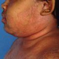
Balanitis describes inflammation of the glans penis and posthitis means inflammation of the prepuce. It can be of diverse etiology, infective (most common being candidial but occasionally can be herpetic as well) (Figs. 15.1 and 15.2) and non-infective. Balanitis is common in uncircumcised men as a result of poorer hygiene and aeration or because of irritation by smegma. Many cases of balanitis seen in practice are a simple intertrigo. Topical corticosteroid alone or in combination with antifungal is a common prescription but simple measures and patient education should be the first priority. Rapid resolution can be achieved by advising the patient to keep his foreskin retracted if possible, having advised him of the risk of paraphimosis. Saline baths are also useful and talcum powders are helpful in drying the area. This advice is simple, but compliance may be challenging. Many patients will present having tried steroid-antifungal creams, often obtained over the counter. Such cases usually come with relapse. The simple measures have a more durable effect [14].
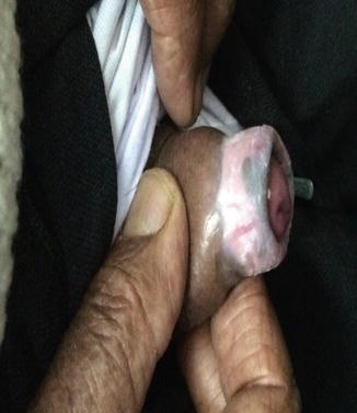
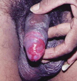
Fig. 15.1
Candidial balanoposthitis in a diabetic patient treated with TS
Fig. 15.2
Herpetic balanitis treated with TS
15.2.3.2 Plasma Cell Balanitis/Zoon’s Balanitis
It is an idiopathic, benign disorder of uncircumcised male genitalia in middle aged. It is a chronic, reactive, principally irritant, mucositis brought about by dysfunctional prepuce with friction playing a part (dorsal aspect of the glans). It is characterized by a circumscribed, persistent moist plaque with a shiny smooth surface on the glans penis and has minute red specks called ‘Cayenne pepper spots’. The keratinized penile shaft and prepuce are spared. Treatment includes moderately potent steroid preparation with or without topical antifungal agents, tacrolimus, CO2 laser and copper vapor LASER. Circumcision is curative.
Ram Chander et al. reported a case of a 70-year-old, married, sexually inactive, uncircumcised male, with biopsy proven plasma cell balanitis with no signs of malignancy. The patient was instructed to apply 0.03% tacrolimus ointment twice daily. Improvement of the lesion was observed after 2 weeks of treatment. The treatment was continued for four more weeks and then tapered over the next 2 weeks. No side effects were observed [15] (Fig. 15.3).
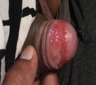
Fig. 15.3
Zoon’s balanitis
15.2.3.3 Lichen Sclerosus et Atrophicus
Lichen sclerosus et atrophicus (LSA) is inherently itchy condition progressively affecting the prepuce, glans and meatus with propensity for atrophy and penile squamous cell carcinoma. Control of pruritus with antihistamines (sedatives) is the key to offer symptomatic relief and arrest progression. Super potent corticosteroid ointment like clobetasol propionate prescribed in a tapering manner over a period of 10–12 weeks controls pruritus. Role of emollient cannot be over emphasized. Secondary bacterial and candidial infection should be treated. Other treatment reported to be efficacious include testosterone ointment, oral stanozolol, acitretin, isotretinoin, ACTH, liquid nitrogen cryotherapy, CO2 laser. Surgical interventions like circumcision and plastic repair are indicated in recalcitrant cases [16] (Fig. 15.4).
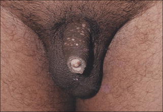
Fig. 15.4
Lichen sclerosus atrophicus with meatal narrowing
15.2.3.4 Balanitis Xerotica Obliterans
It is a form of LSA which occurs in uncircumcised men. Whitish smooth atrophic plaques on glans and prepuce are seen with rarely bullae at affected site. External meatus involvement is noticed in 50% cases with consequent stricture formation. SCC may develop in plaques in long standing cases. Steroid creams have been shown to limit the progression of the disease but do not offer a cure in the majority of cases. Studies have shown that applying a potent topical steroid improves BXO in the histologically early and intermediate stages of disease and may inhibit further worsening in the late stages but do not offer a cure in the majority of cases [17].
15.2.3.5 Pseudoepitheliomatous Micaceous and Keratotic Balanitis
It is a rare penile condition which is seen mainly in elderly over 60 years. It is characterized by an initial plaque stage, late tumor stage followed by verrucous carcinoma and transformation to SCC and metastasis. Coronal balanitis with silvery white appearance, mica-like and keratotic horny masses on the glans is seen. The choice of treatment is generally guided by the stage of the disease. Topical measures include 5-FU, podophyllin resin, potent topical steroids and physical measures are cryotherapy, radiotherapy and surgical excision (Fig. 15.5a–c).
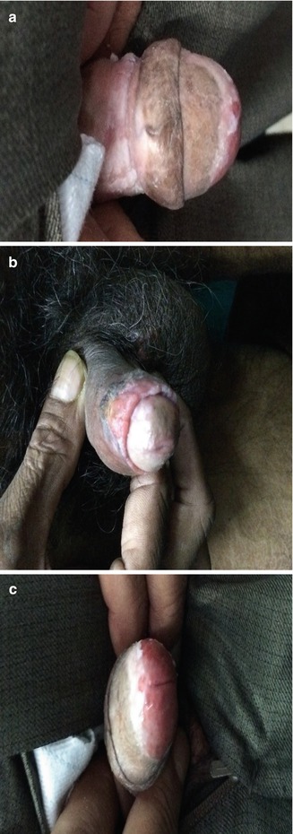
Fig. 15.5
(a–c) Pseudoepitheliomatous micaceous and keratotic balanitis
15.2.4 Females
15.2.4.1 Plasma Cell Vulvitis
It represents a reaction pattern to an inflammatory condition causing intractable vulvar pruritus. Treatment modalities are topical corticosteroid, lignocaine and misoprostol. Çelik A et al. reported a case of plasma cell vulvitis for which topical clobetasol 17-dipropionate cream 0.05% was applied twice daily. Three weeks after starting the treatment with the topical steroids, 50% of complaints had resolved and the lesions significantly improved. After 3 months, all the symptoms and signs were relieved.
15.2.4.2 Vulvar Lichen Sclerosus Atrophicus
Vulvar lichen sclerosus atrophicus (VLS) is an intensely pruritic chronic inflammatory dermatoses resulting into disfiguring sequelae and having malignant potential. Most commonly occurs on the vulva and around the anus with ivory-white lesions appearing like figure of eight that may be shiny. It may be asymptomatic in some cases. Treatment options include potent topical corticosteroids and tacrolimus.
As per the study done by Renaud-Vilmer C et al., prolonged treatment with 0.05% clobetasol propionate ointment resulted in improvement in women older than 70 years and complete regression in younger women with recurrence. Lifelong follow-up is recommended in all cases [18] (Fig. 15.6).
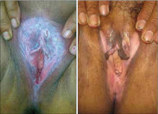
Fig. 15.6
Vulvar lichen sclerosus
15.2.5 Genital Pruritus
Genital pruritus without apparent skin lesions is empirically treated with topical steroids but common causes should be ruled out. Atopic diathesis is to be considered as an endogenous cause. Exogenous causes include irritation and allergy. Genital allergy can be related to sexual activity, non-sexual causes like topical medication and use of genital hygiene products or even consort contact dermatitis [19].
Related posts:
 Evolution and Development of Topical Corticosteroids
Topical Corticosteroid Addiction
Topical Side Effects of Topical Corticosteroids
Evolution and Development of Topical Corticosteroids
Topical Corticosteroid Addiction
Topical Side Effects of Topical Corticosteroids
 Dermatological Indications and Usage of Topical Corticosteroid
 Topical Corticosteroid Modified Superficial Dermatophytosis: Morphological Patterns
 Topical Corticosteroid Abuse: Southeast Asia Perspective
Stay updated, free articles. Join our Telegram channel

Full access? Get Clinical Tree


