Indications
Relative contraindications
Resections over 20 % of breast volume
Extensive tumors located in medial regions
Macromastia
Low-volume breasts, and without ptosis
Severe ptosis and asymmetry
Previously irradiated breasts
Need for large skin resections inside the mammoplasty area
Large skin resections beyond the mammoplasty area
Central, medial, and inferior tumors
Tobacco addiction and uncontrolled diabetes
Previous plastic surgery of the breast
Exaggerated patient expectations with aesthetic results
11.3 Preoperative Planning
It is essential that the choice of the technique for oncoplastic surgery in breast-conserving surgery depends on elements related to the tumor location, size, and multifocality/muticentricity, characteristics of the breast, and clinical evaluation of the patient. Although the only significant element mentioned as an aesthetic risk in breast-conserving surgery in the Cochrane evaluation was a mammary resection volume over 20 %, in clinical practice there are other individualized risk factors that should be observed [23]:
Tumor size/breast size
Multicentricity and multifocality
Location of tumor and proximity to skin
Distance between the tumor and the nipple–areola complex (NAC)
Previous and future radiotherapy
Previous mammoplasty
Volume and shape of the breast
Level of mammary ptosis and breast asymmetry.
Liposubstitution level.
In some circumstances, associated clinical conditions may influence the choice of the most appropriate technique. Diabetic patients, obesity, tobacco addicts, those with collagen diseases, and those above 70 years old are subject to risks concerning unsatisfactory aesthetic results and skin-healing complications are greater. Major resections and wide NAC dislocations may bring additional risks of fat necrosis and partial or total NAC losses [23].
The ideal location for a tumor is within the wide resection area, or inside the mammoplasty area. When the tumor is close to the skin and outside this area, the oncoplastic procedure may be more complex and it may require combined techniques, whose results are not always satisfactory. In such cases, skin-sparing or nipple-sparing mastectomy should be considered as an option, as well as in cases where a major resection of the skin is needed. Flaps such as latissimus dorsi flap, which has a different color and texture compared with the breast, usually lead to unsatisfactory results, and therefore should be considered as an exception [23].
A high-volume breast with severe ptosis allows surgical procedures with wider margins and usually leads to more satisfactory results. Patients with macromastia represent a formal indication for oncoplastic surgery owing to better radiotherapy planning. In cases of previous breast augmentation plastic surgery, it is necessary to take into consideration that the breast volume is not the real one, and consequently some considerable deformities may result. The biggest problem concerning oncoplastic surgery is dealing with young patients, with a conic breast, without mammary ptosis, and with low or medium volume. In such cases, according to the location or tumor size, local flaps offer little chance of good results, so skin-sparing or nipple-sparing mastectomy with immediate reconstruction may be the best choice [23].
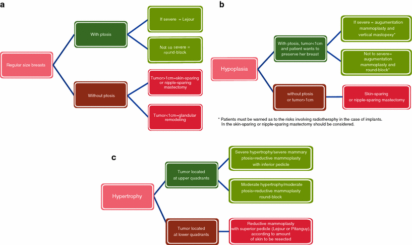
The decision for oncoplastic surgery is based on oncologic and aesthetic concepts and principles, so a structured guideline is not possible to assist in all cases with all involved variables, but it can help the decision-making process. Basically, the flowchart for planning oncoplastic surgery should take into consideration the features of the patient’s breast and the tumor size (Figs. 11.1 and 11.2) [23].
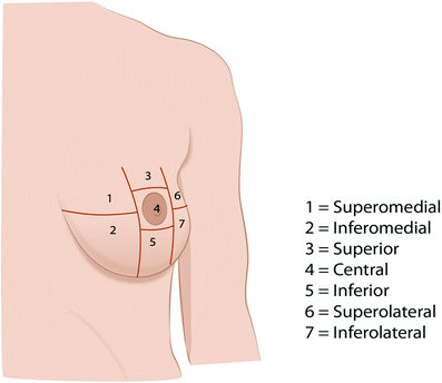

Fig. 11.1
Breast quadrants for oncoplastic surgery
11.4 Immediate Partial Breast Reconstruction Techniques
11.4.1 Class I Techniques
11.4.1.1 Planning for Glandular Flaps
This class I technique consists of moving glandular flaps around the defect caused by classic quadrantectomy or lumpectomy resections, in an attempt to cover it completely. It is preferentially indicated for premenopausal patients, when the glandular component of the breast is greater, therefore reducing the risk of liponecrosis in the postoperative period. This technique is also indicated in cases of tumors located in the upper quadrants, where the mammary gland is less thick; and even if there is a small filling defect, such a defect is not so visible. The opposite effect happens in the lower quadrants, where the mammary gland thickness is more important to consider, and if an adapted technique is not applied, the resulting aesthetic defect may be disastrous. Glandular reshaping in lower portions of the breast is possible for small tumors, and in a vertical or oblique way. Planning for the position of the incisions should consider the quadrant of tumor location (Figs. 11.3 and 11.4).
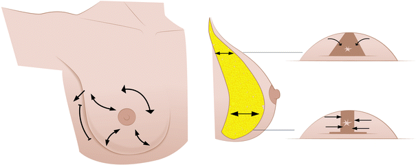
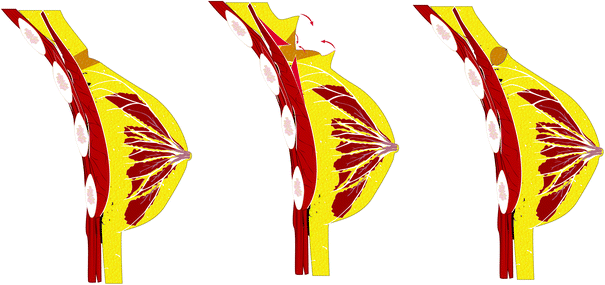

Fig. 11.3
Class I glandular flaps: skin incisions and plan for glandular reshaping

Fig. 11.4
Class I glandular reshaping for superior quadrants
11.4.1.2 Planning for Central Quadrant Techniques
This represented a great innovation in the early days of BCT, as up to some years ago having a retroareolar neoplasia was synonymous of mastectomy. Immediate breast reconstruction techniques for central quadrant resections may differ according to breast volume, level of ptosis, and shape of ptosis (either vertical or lateral). For a breast without ptosis or with slight ptosis, it is possible to use the glandular suture in a tobacco pouch. Two or three layers of glandular suture in a tobacco pouch allow one to obtain the central projection of the mammary cone, and the cutaneous suture also in a tobacco pouch would produce a residual scar within the area where the future areola will be reconstructed, therefore causing the scar to disappear almost completely (Fig. 11.5). The disadvantage of this technique is that there is no good connection with the cutaneous edge and consequently there might be delay in the healing process. A cutaneous–glandular flap can be an alternative and can also result in a good outcome in these cases.
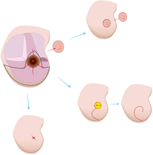

Fig. 11.5
Central quadrant plan for reconstruction with the tobacco purse string technique and a cutaneous–glandular flap
For large breasts with oblique ptosis, it is possible to use a technique described by Galimberti et al. [24], derived from the reductive mammoplasty technique based on the rotation of the inferolateral glandular pedicle, preserving a cutaneous island that replaces the areolomammillary complex (Fig. 11.6). This might be the first oncoplastic technique described in the literature, as it was a direct adaptation of the plastic surgery technique to BCT.
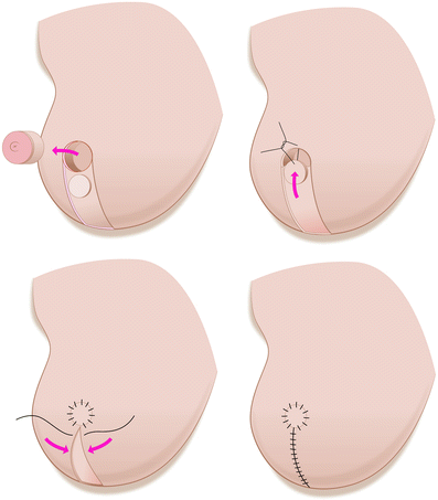

Fig. 11.6
Plan for Grisotti’s flap for the central quadrant
11.4.2 Class II Techniques
11.4.2.1 Planning for Periareolar Techniques
These class II techniques are inspired by reductive mammoplasty techniques proposed by Góes [25] and Benelli [26], in which a major glandular cutaneous undermining procedure for remodeling through a periareolar scar is performed. It is indicated for small or medium-volume breasts with little or average ptosis. The great advantage of these techniques is mainly oncologic, as they allow for a tumorectomy or either a simple or a double quadrantectomy in any part of the breast, except for the central quadrant.
Stay updated, free articles. Join our Telegram channel

Full access? Get Clinical Tree


