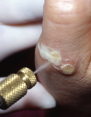
Chapter 23 This involves the destruction of tissues by extreme cold (Box 23.1). The tissue is frozen to subzero temperatures, which is then followed by sloughing of dead tissue. Several mechanisms are involved including the osmotic effects of intracellular water leaving the cell and causing dehydration, intracellular ice formation disrupting the cell membrane and ischaemic damage due to freezing of vessels. Liquid nitrogen is most commonly employed, although various freezing agents are available such as solid carbon dioxide, nitrous oxide and a mixture of dimethyl ether and propane. Unless otherwise stated the rest of this section relates to liquid nitrogen cryotherapy. The low temperature of liquid nitrogen (−196 °C), ease of storage and relative low cost make it an effective and convenient cryogen. However, its low temperature also results in rapid evaporation and therefore it should be stored carefully in an adequately ventilated area and preferably in a pressurised container. The liquid nitrogen is best applied as a spray using a canister (Figure 23.1). Figure 23.1 Cryotherapy. An alternative method is to use a cotton bud that is immersed in liquid nitrogen and then applied to the lesion being treated, using moderate pressure until frozen. More than one application may be needed. A fresh cotton bud should be used for each patient to diminish the risk of transferring human papillomavirus. However, with this method there in an increase in temperature partly due to poor thermal capacity of the cotton and also warming when the cotton tip is transferred from the liquid nitrogen container to the patient’s skin. The freeze time is important, and will vary according to the lesion being treated. Freeze time is counted from the moment the entire lesion becomes frozen white rather than simply from when spraying begins. Once spraying is complete, the rate of thawing of the tissue is an important factor as more tissue destruction occurs with rapid freezing and slow thawing. The ‘freeze–thaw’ cycle may be repeated to increase the degree of damage and the additional freeze has a greater penetration due to improved cold conductivity of the previously frozen tissue. Freeze times and the number of freeze–thaw cycles depend on the type of lesion (i.e. whether benign or malignant) as well as the size and thickness. Cryotherapy is usually initiated on the basis of a clinical diagnosis without prior histological confirmation, and therefore, the clinician must be confident of the diagnosis. If there is any diagnostic doubt, consider a biopsy first or alternative treatment modality where histology can also be obtained. The following lesions are frequently treated with cryotherapy. A single freeze lasting 10–30 s per treatment, which includes a 1–2 mm margin of normal skin, is often sufficient, although a double freeze–thaw cycle may improve clearance, particularly for thicker warts. Usually several treatments at 2–3-week intervals are necessary and cryotherapy may be combined with topical therapies such as salicylic acid preparations for increased efficacy. Paring down the wart with a blade before cryotherapy can also be helpful. A single freeze of between 5 and 20 s including a 1- to 2-mm margin of normal skin should be effective for most lesions. A frozen lesion once thawed for a few seconds can also be curetted off. Larger, thicker lesions may require prolonged freezing or repeat freeze–thaw cycles, thereby increasing pain and inflammation. In these circumstances, it may be better to curette and gently cauterise the area. A single freeze of 5–10 s may be sufficient and it is helpful to stabilise the skin tag with metal forceps so that the liquid nitrogen is sprayed obliquely, avoiding non-lesional skin. An alternative method is to treat by compression with artery forceps dipped in liquid nitrogen. A single freeze of between 5 and 15 s including a 1- to 2-mm margin of normal skin is advised. When necessary, lifting away hard keratin to expose the underlying abnormal epithelium makes the freezing more effective. Rarely, a double freeze–thaw cycle may be needed but be aware that a lesion which does not respond to cryotherapy may be a squamous cell carcinoma. This is an intraepidermal (in situ) form of squamous cell carcinoma, which can be effectively treated with a single freeze of up to 30 s including a 1- to 2-mm margin of normal skin. Again, a biopsy is necessary should there be any doubt about the diagnosis, and follow-up is essential to make sure the lesion has cleared and is not progressing. If cryotherapy is to be employed, it is best limited to the treatment of the superficial type of basal cell carcinoma (BCC), when lesions are primary (i.e. previously untreated), small (<1 cm in diameter) and well defined. The cure rate for other types of BCC is inferior with cryotherapy than with other forms of treatment such as excision. Two cycles of freezing lasting between 20 and 30 s, including a 3-mm rim of clinically normal skin, with a thaw time of 2 min is effective. This term describes the use of electricity to cause thermal tissue destruction. There are two main forms of treatment: electrocautery and electrodessication.
Practical Procedures and Skin Surgery
OVERVIEW
Cryotherapy
Box 23.1 Cryotherapy—practical points
Application technique

Risks and precautions
Skin lesions suitable for freezing
Viral warts
Seborrhoeic keratoses
Papillomas and skin tags
Actinic keratosis
Bowen’s disease
Basal cell carcinoma
Electrosurgery
Stay updated, free articles. Join our Telegram channel

Full access? Get Clinical Tree


