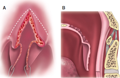
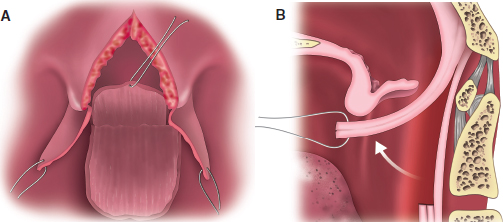
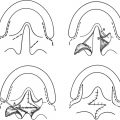
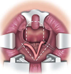
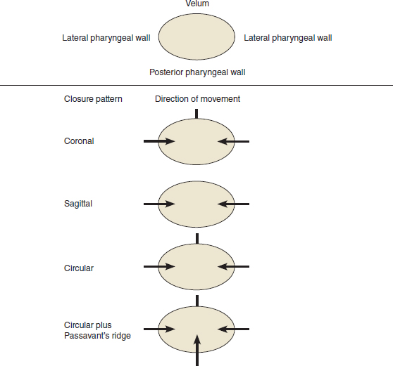
59 ○ One in three children may develop velopharyngeal dysfunction after cleft palate repair. ○ The decision to treat velopharyngeal dysfunction depends on many factors, including patient age, duration of the velopharyngeal dysfunction, the cause of velopharyngeal dysfunction (congenital versus acquired, structural versus idiopathic, neuromuscular), the severity of the dysfunction, the intellectual capacity of the patient, comorbidities, and the presence of mitigating factors such as obstructive sleep apnea. ○ Multidisciplinary assessment should include evaluation by a speech-language pathologist. Subjective speech assessments should document speech intelligibility, resonance, articulation, and nasal air escape. Investigations such as nasendoscopy, nasometry and multiview video fluoroscopy will help delineate the degree and cause of velopharyngeal dysfunction. ○ Surgical procedures to correct velopharyngeal dysfunction can be categorized anatomically and physiologically and should be tailored to the needs of the patient. ○ Superiorly based pharyngeal flaps have a good success rate for correcting velopharyngeal insufficiency, but the patient should be monitored for potential complications such as obstructive sleep apnea. The inability to articulate and speak well may severely stigmatize a child and carries significant psychosocial implications. An estimated 10% to 36% of children who have had cleft palate repair suffer from velopharyngeal dysfunction.1–6 The inability of the velopharyngeal sphincter to isolate the nasopharynx from the oropharynx during speech leads to velopharyngeal insufficiency (VPI). The literature is full of inconsistencies in terminology used to describe disorders of the velopharyngeal valve. The terms insufficiency, inadequacy, incompetency, or dysfunction are often used interchangeably and may or may not be used correctly to differentiate underlying etiology. For the purpose of this chapter, we have elected to use the term velopharyngeal insufficiency, because it is the most common term used to describe anatomic or structural etiologies, such as in cleft palate, and is the term most commonly used in our practice at SickKids Hospital in Toronto. The characteristics of VPI may include hypernasal resonance; nasal air emission; nasal or facial grimacing; nasal turbulence; inadequate intraoral air pressure resulting in weak, nasalized, or absent consonants; and compensatory articulation substitutions. Compensatory articulations develop as the result of consonants produced at the level of the pharynx or larynx in an attempt to shape the airstream more posterior in the vocal tract. The speech sequelae of VPI can have a profound effect on an individual’s ability to be understood (speech intelligibility), jeopardizing social integration and peer acceptance. Fortunately, a combined approach of surgery and speech therapy can produce an excellent prognosis for most patients. Surgical techniques to correct VPI may be characterized as static (flaps) or dynamic (sphincter techniques). As a static procedure, the posterior pharyngeal flap represents the most common surgical technique used to treat VPI after cleft palate repair.1,2 In a recent survey of cleft palate and craniofacial professionals, 52.9% reported that the pharyngeal flap was the most common procedure used in their centers to correct VPI.7 However, the correction of speech problems using this technique is one of the least physiologic surgical procedures performed in the cleft armamentarium and can incur significant morbidity. This chapter describes the technique and application of posterior pharyngeal flaps in the management of VPI in patients with clefts. Other chapters discuss anatomic concerns, speech evaluation, and other surgical techniques for velopharyngeal insufficiency correction. In general, a single operation is not likely to correct all cases of velopharyngeal insufficiency because of the heterogeneity of underlying medical conditions, intellectual capacity, and degree and type of attempted closure patterns in patients with this condition. Optimal results are obtained by customizing the procedure to fit the patient’s needs based on careful preoperative assessment. Gustav Passavant8 is credited as being the first to attempt surgical correction of velopharyngeal insufficiency by suturing the posterior border of the soft palate to the posterior pharyngeal wall in 1865. The inferiorly based pharyngeal flap was originally described by Karl Schoenborn9 in 1875 and was first performed on a 17-year-old girl with an unrepaired cleft of the hard and soft palate. A fistula that developed between the left side of the flap and the palate required a second von Langenbeck procedure for complete healing, but Schoenborn was impressed that the patient’s speech was clear and understandable as soon as healing had taken place. Schoenborn eventually switched to a superiorly based flap, because he felt that suturing the inferiorly based flap was technically difficult as a result of the fragile adenoid tissue. He also advocated dividing the flap if the patient maintained acceptable speech for some years, but whether this was common practice is unknown. In 1924, Wolfgang Rosenthal10 combined the inferiorly based pharyngeal flap with a von Langenbeck cleft palate closure for the first time and claimed perfect speech results. The pharyngeal flap was popularized in the United States by Padgett11 in the 1930s and has been modified by many authors. Length and width of the flap, donor site closure, techniques of attachment, and insetting all underwent various degrees of adjustment in the 1950s and 1960s. However, the key contributions to improving the superior pharyngeal flap technique have consisted of lining the raw surface of the flap, lateral port control, and tailoring the size to suit the patient’s needs using data collected from preoperative videofluoroscopy and nasendoscopy. According to Millard,12 splitting the soft palate to improve exposure and reflecting nasal mucosal flaps to cover the raw undersurface of the pharyngeal flap was suggested by Blackfield in 1963. Further attempts to provide lining to the superiorly based flap included, in 1975, Isshiki and Morimoto’s technique13 of folding it back on itself in the hopes that it would minimize postoperative scar contracture and shrinkage. However, it was Michael Hogan’s concept14 of attempting to provide control of the lateral port size initially in 1971 that led to the most commonly used method for flap lining (Fig. 59-1). Previous work had demonstrated that oropharyngeal pressure began to decrease when the size of the lateral pharyngeal port was greater than 10 mm2 and was greatly impaired when the port size was 20 mm2. This led to significant nasal air emission and hypernasality; Hogan concluded that the 20 mm2 area represented the threshold of velopharyngeal incompetence. He decided to modify the superior pharyngeal flap to improve control of the lateral port size by using a catheter with an external diameter of 4 mm2 to establish the port size during insetting of the flap. By his publication in 1973, he believed he had created a total surface area of the two ports of about 25 mm2 and claimed a success rate of 97% in a series of 93 patients. However, he was aware of the risk of creating significant hyponasality and an absence of nasal respiration, snoring, and increased mucous production. Hogan recommended reopening the ports in adults but being cautious of doing so in the growing child. He described lining the pharyngeal flap with nasal mucosal flaps to maintain flap size and prevent shrinkage. His innovative modifications continue to receive wide acceptance. Fig. 59-1 Setup for the Hogan modification of the superior flap technique. The soft palate has been split down the middle and retraction sutures placed. Nasal flaps are marked out and may be triangular or rectangular in shape. The superior flap is marked out on the posterior pharyngeal wall with the upper extent at the adenoid pad, allowing insetting at the level of the soft palate. Shprintzen et al15 and several others contributed in important ways to improving success and decreasing morbidity by suggesting that the flap be tailored to the needs of the patient based on preoperative nasendoscopy and videofluoroscopy.1 Modifications of this versatile flap continue. The posterior pharyngeal wall is an ideal donor site and provides unscarred, well-vascularized, nonviolated expendable tissue with minimal long-term donor-site morbidity. The donor site consists of the region of the pharynx bounded superiorly by the adenoid pads and extending inferiorly to the region of the third cervical body. The lateral pharyngeal walls define the lateral extent. Flaps designed in the pharynx may be classified as static or dynamic, inferiorly based, superiorly based, or laterally based (Table 59-1). This chapter addresses static posterior pharyngeal flaps. These are musculomucosal flaps that incorporate segments of the palatopharyngeus and superior constrictor muscles. Flaps based superiorly have the advantage that they can be made longer than inferiorly based flaps, in which the length is limited by the presence of the adenoid pad. Regardless of the location of the flap base, the aim of the pharyngeal flap procedure is to create a central subtotal velopharyngeal obstruction with two small lateral ports remaining for nasal air flow. Table 59-1 Classification of Pharyngeal Flaps Although the anatomy of the nasopharynx has been covered in previous chapters, some specific points are worth noting. The muscles of the posterior pharyngeal wall consist of the superior pharyngeal constrictor, which is a curved quadrilateral sheet of muscle that encompasses the nasopharynx and upper oropharynx and is the thinnest of the three constrictor muscles. It originates from the posterior pharyngeal raphe and runs laterally and anteriorly to insert into the palate and hamulus superiorly and the pterygomandibular raphe and mandible inferiorly. It is innervated by the cranial part of the accessory nerve from the pharyngeal plexus. Contraction of the superior constrictor muscle results in medial and anterior movement of the lateral pharyngeal walls. The palatopharyngeus muscle forms part of the palatopharyngeal arch and is composed of two fasciculi that lie in the same plane but are separated by the levator veli palatini. These two layers unite at the posterolateral border of the soft palate and run downward and lateral to form an internal incomplete muscle layer in the pharyngeal wall. It is also innervated by the cranial part of the accessory nerve from the pharyngeal plexus. Passavant’s ridge is a thickened band of muscle at or below the level of the soft palate. This muscular band is visible when the soft palate is elevated during pneumatic activities that use positive air pressure, such as speech, whistling, and blowing. The size of Passavant’s ridge can vary according to the type of velar activity. It can be seen in both normal speakers and those with velopharyngeal dysfunction. It does not appear to be correlated to gap size or type of cleft palate. The vertical location of the ridge is variable among individuals but is consistent within an individual. Controversy exists as to whether this represents a separate distinct muscle (Passavant’s muscle) or whether it is a part of the superior constrictor and palatopharyngeus muscle.16 This is also the site of the transition from the columnar ciliated respiratory epithelium of the nasopharynx to the stratified squamous epithelium of the oropharynx. Lateral wall movement of the pharynx results from contraction of the superior constrictor and palatopharyngeus muscles that form hemisphincters, causing medial displacement of the lateral pharyngeal walls. Elevation of a pharyngeal flap may affect lateral pharyngeal wall function because of the disruption of the transverse fibers of both of these muscles by the vertical incisions of the flap. Although Zwitman17 documented reduced lateral wall movement after posterior pharyngeal flap surgery, this finding was not repeated in subsequent studies, suggesting considerable ability to compensate.15,18 Anatomic studies of the blood supply to inferiorly and superiorly based pharyngeal flaps were performed by Mercer and MacCarthy19 using radiopaque injection techniques in cadavers. The study documented the anatomy of the ascending pharyngeal artery, arising as a branch of the external carotid artery, as the primary blood supply to the pharynx. Several interesting findings were reported. The first was a dehiscence of the superior constrictor in the midline over a distance of 8 mm from the cranial base. This region is the arch of C1 or the lower pole of the adenoid pad and implies that the mucosa is attached directly to the pharyngobasilar fascia. Therefore a superiorly based flap would not be likely to have functional muscle in its base unless the base was placed lower than the first cervical vertebrae. Second, raising a superiorly based flap divides the pharyngeal branches of the ascending pharyngeal artery that run under the prevertebral fascia and supply the overlying muscle and mucosa. No axial vessels were detected in the flap. This suggests that the superiorly based flap has a random pattern, and the guidelines of length-to-width ratios should be recognized. Ischemia may therefore be an additional reason why these flaps are known to shrink and form tubes over time, although muscle denervation is also a recognized factor. Flap shrinkage has been documented to be about 50% of its original width.20 Ischemia may also contribute to flap dehiscence, which has been reported in up to 7% of cases.19,21,22 Finally, inferiorly based and laterally based flaps are likely to contain axial vessels that allow flaps with a length-to-width ratio greater than 2:1 to be elevated. The popularity of such flaps has been limited because of limitations in flap length and tethering of the palate low in the pharynx. Indications for treatment of VPI should be derived from both subjective and objective data. Subjective speech assessments should document speech intelligibility, resonance, articulation, and nasal air escape. Objective measures of nasality using various speech stimuli include the nasalance score, which is a ratio of the nasal acoustic output relative to oral plus nasal acoustic output and is expressed as a percentage. This score is matched against age- and dialect-appropriate normal values. Nasendoscopy provides direct visualization of the soft palate and surrounding velopharyngeal structures during speech and nonspeech activities. The type of closure pattern, symmetry of movement, degree of closure, palatal morphology, presence of tonsils and adenoids, and pharyngeal pulsations should all be documented. The velopharyngeal closure rating (out of 1.0 in 0.1 increments) is based on the fraction of the diameter of the velopharyngeal port that is closed off during attempted sphincter closing.23 Multiview videofluoroscopy may also be used for determining the defect size and closure pattern but carries the risk of radiation exposure; at the Hospital for Sick Children in Toronto, it is reserved for noncompliant children in whom nasendosopy is not tolerated or when lateral-view imaging (which cannot be obtained using nasendoscopy) is needed. The preoperative closure pattern and the selection of best surgical procedure has been a debated topic in the literature.24 Armour et al25 from the Hospital for Sick Children in Toronto hypothesized that the closure pattern of the velopharyngeal sphincter was an important determinant in selecting the proper operation for children with VPI. Closure patterns may be classified into coronal (good velar movement), sagittal (good lateral wall movement), or circular (good velar and lateral wall movement) plus or minus a contribution from Passavant’s ridge26 (Fig. 59-2). In a retrospective correlation, they demonstrated that the preoperative velopharyngeal closure pattern was a significant factor in predicting the effectiveness of superiorly based pharyngeal flaps in correcting the hypernasality associated with VPI. As a static procedure, success of the superiorly based flap depends on lateral port closure because of the medial movement of the lateral pharyngeal walls. This study demonstrated that noncoronal (sagittal and circular) closure patterns were satisfactorily addressed by the superiorly based flap technique. A coronal closure pattern was associated with poor lateral pharyngeal wall movement resulting in a lower success rate. Nabi et al24 found that preoperative circular closure patterns had better posttreatment nasalance scores versus coronal patterns; however, the reverse was true when they looked at longer term results for audible nasal air emission and compensatory articulation perceptual speech scores. Other studies have failed to show an advantage in speech outcomes between coronal and circular closure patterns in patients who have pharyngeal flap surgery and suggest improved lateral pharyngeal wall movement after the procedure.27,28 Shprintzen et al29 advocated for tailor-made flaps based on the degree of preoperative lateral wall adduction. Fig. 59-2 Anatomy of the velopharyngeal port and closure patterns. The decision to treat VPI depends on many factors, including patient age, duration of VPI, cause of VPI (congenital versus acquired, structural versus idiopathic, neuromuscular) severity of VPI, intellectual capacity of the patient, comorbidities, and the presence of mitigating factors such as obstructive sleep apnea. Nonsurgical therapies include prosthetic management with speech bulbs and palatal lift devices, but in general these are not well tolerated in children. At the Hospital for Sick Children in Toronto, the spectrum of surgical management of VPI consists of superiorly based pharyngeal flaps, sphincter pharyngoplasty, and palatal lengthening techniques (Furlow double-opposing Z-plasty or pushback palatoplasty). Rarely, augmentation techniques (fat grafting) are used. Inferiorly and transversely based flaps are not routinely performed. Table 59-2 Treatment Indications for Velopharyngeal Insufficiency These recommendations are based on findings at nasendoscopy. Indications for superiorly based pharyngeal flaps are VPI associated with poor palatal motion, a velopharyngeal gap detected on nasendoscopy or multiview videofluoroscopy, and the presence of some lateral wall movement. Palatal lengthening techniques may be applied when velopharyngeal closure is marginal (0.7 closure rating or greater), an overt or occult submucous cleft palate has been detected, or when aberrant vessels in the posterior pharyngeal wall precludes safe elevation of a pharyngeal flap. Poor lateral wall movement (coronal closure) may be an indication for sphincter pharyngoplasty (Table 59-2). Regardless of the surgical technique, postoperative speech therapy must be explained to patients and families and they must understand the limitations of surgery for VPI management to be successful. Neither the inferiorly nor superiorly based flap has been proven superior to the other. The inferiorly based flap may have an advantage in terms of vascularity but has limitations because of its length, the inability to suture the flap tip, and the low position of the base in the pharynx. The superiorly based flap may be lengthened and inset at the level of the soft palate, but it may be lacking as robust a vasculature. Previous studies by Skoog,30 Hamlen,31 Whitaker et al,32 and Karling et al28 found no significant differences in postoperative speech, hearing, complications, or length of stay between those patients who had a superiorly based compared with an inferiorly based flap. The diagnosis of VPI is not usually established until the child is about 4 years of age. This allows the language and articulation systems a chance to mature and to benefit from speech therapy, which should be instituted as soon as a speech disorder is detected. In some cases, improvements in velopharyngeal valving occur as articulation skills improve. This is particularly true when compensatory articulation substitutions can be remediated. In addition, patient cooperation for visualization techniques (nasendoscopy and multiview videofluoroscopy) needs to be considered. Once all necessary data has been obtained, and it is documented that VPI is not going to resolve with only speech therapy, surgical management should be considered. Ideally, surgery should be performed at about 5 years of age. Although some studies have suggested that surgery before 6 years of age may be associated with improved outcomes, others have not shown such an advantage.33–35 All patients in the Cleft Lip and Palate Program at the Hospital for Sick Children in Toronto are seen in a multidisciplinary setting and undergo regular team assessments, including otolaryngology, pediatric dentistry, orthodontics, plastic surgery, speech and language pathology, social work, psychiatry, and audiology at 1, 3, 5, and 9 years, and subsequently depending on the treatment plan for that child. Before pharyngeal flap surgery, an appropriate medical, behavioral, and family history is obtained. Preoperative audiograms are important to rule out conductive hearing loss; when necessary, myringotomy and ventilation tube insertion may be performed with the pharyngeal flap surgery. When appropriate, preoperative cervical vascular imaging is obtained. This issue is addressed in the section on 22q11 microdeletion syndrome. Examination on all patients includes an oral peripheral examination, perceptual judgment of resonance, acoustic analysis (nasometry), and visualization of the velopharyngeal mechanism (multiview videofluoroscopy, nasopharyngoscopy, or both). The patient is placed supine on a headrest with bolsters placed under the shoulders, and the neck is extended. An RAE endotracheal tube is used to secure general anesthesia, placed in the midline, and taped. A Dingman mouth gag is inserted using the largest possible tongue blade; the endotracheal tube must not be compressed when it is opened, nor the posterior pharyngeal wall encroached upon. A small throat pack may be useful. Direct visualization of the pharynx is accompanied by finger palpation to detect aberrant vasculature. A headlight worn by the operating surgeon improves visualization. Prophylactic antibiotics are not routinely administered unless the patient has cardiac issues. Excessive neck extension should be prevented because of the rare but documented occurrence of spontaneous cervical dislocation. Trendelenburg positioning may assist in visualization and improve comfort for the surgeon but will increase venous bleeding. The flap is marked out with the base located at the adenoid pad that correlates to the anterior tubercle of the first cervical vertebrae (see Fig. 59-1). Flap width may vary according to preoperative investigations, but the average flap width ranges between 2 and 3 cm. The width of the flap will depend on lateral pharyngeal wall movement but, in general, wider flaps are used when the lateral pharyngeal wall movement is poor. The flap may be skewed to one side in the presence of abnormal pulsations or when asymmetrical velopharyngeal movement has been preoperatively documented. The length of the flap depends on access and requirements for tension-free insetting. The average flap length is 2.5 to 3 cm, but it may extend inferiorly for up to 4 cm. The tip of the flap has been found to correlate with the second cervical vertebrae. After marking is completed, the posterior pharyngeal wall is infiltrated with lidocaine (Xylocaine) 1% with 1:200,000 epinephrine, and a period of 7 minutes is allowed to ensure vasoconstriction. If the Hogan modification is to be applied, the soft palate is infiltrated with Xylocaine 1% with 1:200,000 epinephrine and, after adequate time for vasoconstriction has passed, the soft palate is split down the middle with a No. 11 blade from the tip of the uvula to the margin of the hard palate (see Fig. 59-1). Retraction on the uvula with 4-0 sutures facilitates this maneuver by significantly aiding exposure for the pharyngeal flap. Triangular or rectangular flaps of nasal mucosa based along the free edge of the soft palate are marked out and raised at a submucosal level using a No. 15 blade or fine dissecting scissors (Fig. 59-3). The posterolateral extent of these flaps determines the size of the lateral port and is critical to the success of the procedure. The length of these flaps should be similar to the depth of the palate. Once these flaps have been elevated, traction sutures are used to keep them out of the field. The lateral incisions of the flap are made with either diathermy or a No. 15 blade, then blunt scissors are used to dissect through the superior constrictor and palatopharyngeus muscles to expose the prevertebral fascia. This is easily identifiable by its white avascular appearance. The flap is undermined in a bipedicle fashion; then, with retraction of the flap in a cranial fashion, sharp cutting scissors may be used to cut the distal end of the flap at the appropriate length. The flap is mobilized completely to its base to allow a tension-free inset (Fig. 59-4). Posterior pharyngeal veins are the most common site for bleeding and are visible on the prevertebral fascia. Donor-site closure is always performed. Closure may improve patient comfort and reduces the risk of postoperative hemorrhage and infection. The muscle and mucosa of the posterior pharyngeal wall is sutured with 3-0 absorbable suture as one layer (Fig. 59-5). A bite of the prevertebral fascia must be picked up in the closure to prevent webbing and narrowing of the pharynx and possibly prevent hematoma formation by eliminating dead space. The cranial end of the donor site should not constrict the flap base during closure.
Posterior Pharyngeal Flaps
Karen W.Y. Wong, Paula G. Klaiman, Christopher R. Forrest
KEY POINTS
EVOLUTION OF POSTERIOR PHARYNGEAL FLAPS
CLASSIFICATION OF POSTERIOR PHARYNGEAL FLAPS
Type
Characteristic
Static
Posteriorly based
Inferior based
Superior based
Laterally based
Dynamic
Sphincter techniques
Hynes
Orticochea
Jackson modification
ANATOMY
INDICATIONS FOR POSTERIOR PHARYNGEAL FLAP SURGERY
Treatment
Indications
Superior pharyngeal flap
Good lateral wall movement
Moderate to large defect
Sphincter pharyngoplasty
Poor lateral wall movement
Furlow (double-opposing Z-plasty) palatoplasty
Small defect
Occult or small submucous cleft palate
Pushback palatoplasty
Moderate to large defect if combined with a superior flap
Small defect if isolated procedure
Nonsurgical (palatal lift device)
Surgical contraindication
Patient request
Training device for borderline or marginal velopharyngeal insufficiency
INFERIORLY VERSUS SUPERIORLY BASED FLAPS
TIMING OF SURGERY
SURGICAL TECHNIQUE
Preoperative Assessment
Patient Preparation
Flap Marking
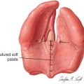
Splitting of the Palate
Posterior Pharyngeal Flap Elevation
Donor-Site Management
Plastic Surgery Key
Fastest Plastic Surgery & Dermatology Insight Engine