17
Posterior Interosseous Syndrome
William B. Geissler
History and Clinical Presentation
A 52-year-old right hand dominant carpenter and avid golfer presented with complaints of pain well localized to the right extensor muscle mass just distal to the elbow joint that had persisted for 6 months. He describes the pain as aching in character with burning that radiates along the anatomic course of the forearm extensor muscles. He has noticed a slowly enlarging mass over the dorsal proximal forearm and complains of weakness in grip strength and diminished endurance. He has no complaints of sensory loss to the hand or forearm. He denies any history of trauma.
Physical Examination
The right forearm measured 2 cm larger than the left in circumference and measured 5 cm distal to the lateral epicondyle. A palpable mass was noted over the dorsal proximal forearm, and the patient was tender to palpation over the radial nerve as it passed through the mobile muscle mass at and just distal to the radial head. Slight tenderness to palpation was noted over the same area to the left forearm but not as severe. Palpation directly over the lateral epicondyle caused mild discomfort, less severe than over the course of the nerve. Full passive flexion of the wrist and fingers with the elbow in extension reproduced the pain. Resisted supination with the extended arm similarly reproduced the pain symptoms. Radial deviation was observed with wrist extension. The patient was able to fully extend the interphalangeal joints, but he could not fully extend the metacarpophalangeal joints beyond 45 degrees.
PEARLS
- Brachioradialis splitting approach provides excellent easy exposure to the radial tunnel for decompression.
- Easier to identify nerve proximally and trace out distally
- Consider posterior approach for mass compressing distal aspect of radial tunnel.
PITFALLS
- Beware of cervical radiculopathy presenting as posterior interosseous syndrome. No sensory symptoms in posterior interosseous syndrome.
- Beware tendon rupture in rheumatoid patients presenting as posterior interosseous nerve syndrome. Treatment is entirely different.
- Revisions usually secondary to incomplete release of nerve through supinator. Compression of posterior interosseous nerve has been reported at distal edge of supinator.
Diagnostic Studies
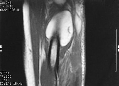
Anteroposterior, lateral, and oblique radiographs of the right elbow did not show any bony abnormalities. An abnormal soft tissue shadow was noted on the lateral radiograph dorsally. Magnetic resonance imaging of the proximal forearm revealed a mass consistent with a lipoma involving the supinator muscle (Fig. 17–1). Electromyographic and nerve conduction studies of the bilateral upper extremities demonstrated denervation of the extensor digitorum communis and extensor carpi ulnaris. The abductor pollicis longus and extensors pollicis longus and brevis were spared.

Figure 17–1. Magnetic resonance image demonstrating a large lipoma overlying the proximal radius. Note the distended supinator muscle.
Differential Diagnosis
Radial tunnel syndrome
Cervical radiculopathy
Lead poisoning
Systemic connective tissue disease
Hysterical wrist drop
Posterior interosseous syndrome
Diagnosis
Posterior Interosseous Syndrome
Radial tunnel syndrome involves compression of the posterior interosseous nerve branch of the radial nerve. Radial tunnel syndrome is defined when there is an absence of muscular involvement and the diagnosis of posterior interosseous syndrome is defined when there is muscular weakness and atrophy in the efferent distribution. The posterior interosseous nerve is not a purely motor nerve. It carries gamma-afferent fibers from the muscles it innervates. This can be demonstrated by the discomfort experienced by squeezing any muscle belly. The posterior interosseous nerve also supplies sensory afferent fibers to the radiocarpal, intercarpal, and carpometacarpal joints. Patients describe an aching, burning discomfort in the mobile extensor wad musculature, which may radiate distally down the forearm.
Stay updated, free articles. Join our Telegram channel

Full access? Get Clinical Tree


