53
Carpometacarpal Joint Injuries: Bennett’s Fractures (Wire Technique)
Michael Jablon
History and Clinical Presentation
A 35-year-old, right-handed dentist injured his left, nondominant thumb in a bicycle accident. His injury occurred when he ran into a cement post. He fractured his right clavicle as well as his left thumb. The thumb fracture was not initially recognized by his surgeon, although initial radiograph reports indicated a “marginated bony fragment adjacent to the medial aspect of the base of the first metacarpal.”
Physical Examination
The left thumb had tenderness at the carpometacarpal joint, with a marked prominence of the first metacarpal base. Skin, two-point discrimination, and Allen’s test were intact.
Diagnostic Studies
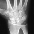
Radiographs of the left hand and thumb carpometacarpal joint indicated the presence of a fracture dislocation. The base of the first metacarpal was fractured intraarticularly and the metacarpal itself was radially and proximally subluxed (Fig. 53–1).
PEARLS
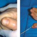
- Once traction, pronation, and abduction have been applied to the thumb for reduction, a K wire may be placed overlying the first metacarpal trapezial level and a fluoroscopic image obtained.
- If the wire is in the appropriate position, draw a line with a marking pen to help as a guide when placing the K wire.
- The transfixing K wire need not cross the fracture site. Strive for perfect anatomic reduction, but a minor step-off may be acceptable.
PITFALLS
- Poor radiographs may delay correct diagnosis. Accurate imaging requires a pronated thumb view to profile the base of the first metacarpal.
- During the surgical approach, care to avoid injury to the superficial radial nerve branches is necessary.
- Prolonged immobilization may lead to loss of mobility and circumduction.
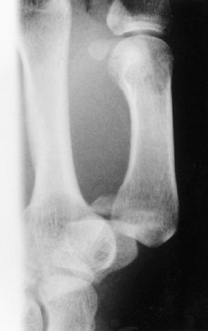
Figure 53–1. Radiograph of patient’s thumb demonstrates small fracture fragment and dorsal radial subluxation of the trapezial metacarpal joint.
Differential Diagnoses
Bennett’s fracture dislocation
Rolando’s fracture dislocation
Metacarpal shaft fractures
Trapezium fractures
Thumb dislocation
Carpometacarpal joint osteoarthritis
Thumb sprain
Pathologic fracture
de Quervain’s tenosynovitis
Diagnosis
Bennett’s Fracture Dislocation
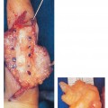
The diagnosis was a Bennett’s fracture and intraarticular fracture of the first metacarpal bone with two fragments; the larger fragment shows subluxation from the deforming pull of the abductor pollicis longus tendon.
The Bennett’s fracture is the most common intraarticular injury involving the carpometacarpal joint of the thumb. Because the abductor pollicis longus tendon acts as a deforming force, stabilization by fixation is necessary. An attempt at closed reduction is recommended by applying traction to the thumb metacarpal with radial abduction and a pronation rotatory torque. A wire is percutaneously driven while holding the thumb in palmar abduction and opposition if reduction is achieved. The pin may be driven into the index metacarpal or trapezium. Always strive for anatomic reduction. However, acceptance of a 2-mm or less articular step-off does not biomechanically predispose a patient to posttraumatic osteoarthritis if the fragments heal in close apposition. The priority of treatment should be reduction of the metacarpal shaft relative to the trapezium and small fragment.
Surgical Management
Related posts:
Stay updated, free articles. Join our Telegram channel

Full access? Get Clinical Tree


