85
Lipoma
Kevin D. Plancher and Michael Bothwell
History and Clinical Presentation
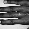
A 40-year-old right hand dominant woman presents with a mass on her right little finger. The mass has grown in size and is now causing functional problems. The patient reports the mass is not painful. She also reports a recent weight gain, corresponding with the enlargement of the mass.
Physical Examination
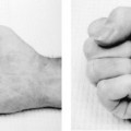
The patient has full range of motion of the little finger and exhibits no strength deficits. The patient has a normal vascular examination. Swelling is present over the hypothenar eminence of the hand. A single round mass is palpated. The patient requests treatment due to functional problems and for cosmetic reasons.
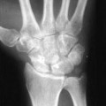
Diagnostic Studies
Radiographic plain films are often useful for revealing the characteristic density of fat, facilitating diagnosis as well as evaluation of the extent of the lesion. Radiographs show an area of decreased density due to increased translucency of the adipose tissue, surrounded by the contrast of adjacent muscle. No calcification was present. Magnetic resonance imaging (MRI) and computed tomography (CT) are helpful if the lesion is deep. Electromyography (EMG) is performed if nerve symptoms are present.
Differential Diagnosis
Ganglions
Synovitis
Rheumatoid nodules
Related posts:

Stay updated, free articles. Join our Telegram channel

Full access? Get Clinical Tree


