46
Lateral Dislocations of the Proximal Interphalangeal Joi
Christopher H. Martin and Steven Z. Glickel
History and Clinical Presentation
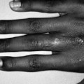
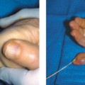
A 43-year-old man caught his left small finger in the coat button of a coworker and the digit was forcefully deviated ulnarward. He felt a snap, and noticed an angular deformity through the proximal interphalangeal (PIP) joint and an inability to fully extend the finger.
Diagnostic Studies
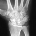
Radiographic evaluation in the emergency room showed a lateral dislocation of the proximal interphalangeal joint (Fig. 46–1). Initial clinical examination showed sensory function to be intact on both sides of the digit, with brisk capillary refill. A digital block was placed and the dislocation was reduced with gentle longitudinal traction. Radiographic examination confirmed a concentric reduction of the PIP joint with a symmetric joint space.
Physical Examination
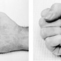
While still anesthetized, the finger was examined to assess stability of the joint and integrity of the extensor system. The patient was able to actively move the digit through a full range of motion. There was no subluxation noted. The integrity of the extensor mechanism and central slip insertion was then tested by having the patient extend the PIP joint against examiner resistance with the metacarpophalangeal (MP) joint flexed. There was full extension with normal strength compared with the uninjured digit.
The digit was splinted with the PIP joint in full extension. The patient was seen weekly for the next 2 weeks with radiographs confirming maintenance of the reduction. After 2 weeks, the digit was buddy taped to the adjacent ring finger and range of motion was begun. Due to some mild laxity in the healing radial collateral ligament, the digit was buddy taped for a total of 4 weeks, after which a full, painless range of motion had been achieved.
PEARLS
- While the patient is under digital block at the time of reduction, assess the integrity of the central slip insertion by resistance to PIP flexion with the MP joint flexed.
- Begin motion as early as patient comfort will allow.
- Assure a concentric reduction: incarceration of the collateral ligaments within the joint will preclude their healing at an appropriate length.
PITFALLS
- Boutonniere deformity
- Stiffness
- Laxity
Differential Diagnosis
Volar PIP rotary dislocation
Lateral PIP dislocation
PIP joint fracture dislocation
PIP joint sprain
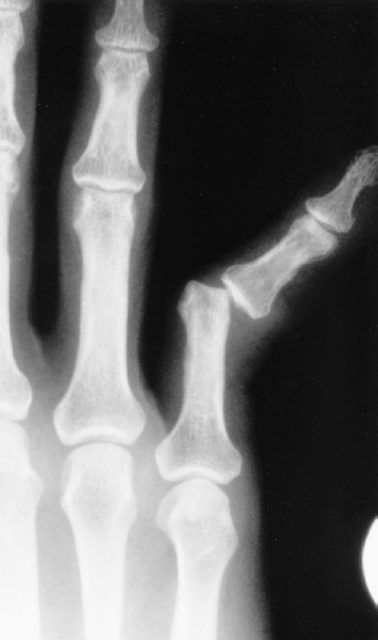
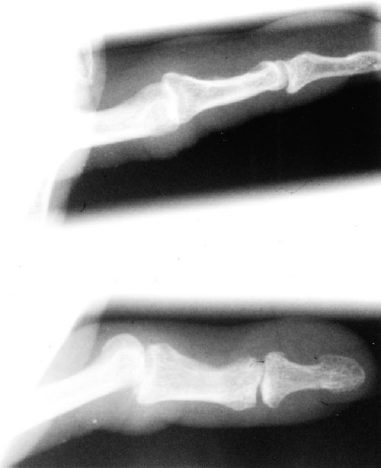
Figure 46–2. Lateral and oblique radiographs of the injured digit show an incongruous PIP joint. The proximal and middle phalanges are in nearly the same plane in the radiograph on the left, suggesting that this is a simple lateral dislocation.
Related posts:

Stay updated, free articles. Join our Telegram channel

Full access? Get Clinical Tree


