Port wine stain (‘nevus flammeus’)


Port wine stain (‘nevus flammeus’)


Andrew C. Krakowski and Lawrence F. Eichenfield


Evidence Levels:  A Double-blind study  B Clinical trial ≥ 20 subjects  C Clinical trial < 20 subjects  D Series ≥ 5 subjects  E Anecdotal case reports


image


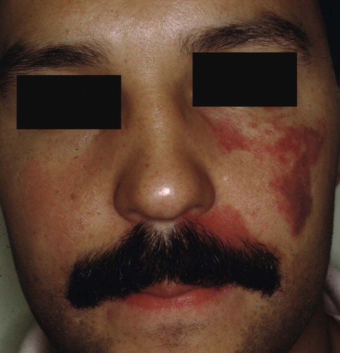
Port wine stains (PWS), also known as ‘nevus flammeus’, are benign capillary malformations of the superficial cutaneous vasculature. These lesions are almost always congenital, though they may be acquired secondary to trauma and, thus, may rarely develop in adolescence or adulthood. The head and neck are sites of predilection, but any part of the integument can be affected. Morphologically, these lesions present as light pink to red patches that typically grow proportionately with the child’s somatic growth. Unlike the salmon patch (also known as nevus simplex, angel kiss), which usually disappears within 1 or 2 years, untreated port wine stains persist throughout a patient’s life and tend to darken with time. Confusing this clinical picture is the fact that PWS may appear to lighten during the first 3 to 6 months of life; this is a physiologic change most likely due to the decrease in blood hemoglobin concentration (typically 15–17 g/dL at birth to a nadir of 8–10 g/dL by age 3 months) and should not be interpreted as a sign of clinical resolution. Skin thickening and the development of surface irregularities (nodules or ‘blebs’) and soft tissue hypertrophy may also occur, especially in the V2 distribution. PWS may also present with an inflammatory component consisting of scaling, excoriations, oozing, and crusting, which resembles an eczematous dermatitis.


While the exact molecular pathogenesis of these capillary malformations has yet to be elucidated, it is believed that complex, localized defects in pathways controlling embryogenesis and angiogenesis play crucial roles. Several specific mutations have recently been identified that may shed light on the possible pathogenesis of capillary malformations. For example, mutations in RASA1, encoding p120-rasGTPase-activating protein (p120-rasGAP), along the Ras/MAPkinase pathway, have been identified in patients with atypical capillary malformations with or without concurrent arteriovenous malformations or arteriovenous fistulas; this protein appears to play a crucial role in controlling proliferation, migration, and cell death in a number of tissues including vascular endothelium. Likewise, developmental endothelial locus-1 (Del-1), an extracellular matrix protein adhered to by human umbilical vein endothelial cells, is another protein being investigated for its potential to induce formation of a vascular plexus with increased number of capillaries. Why certain PWS seem to follow a dermatomal distribution, like the V1 involvement classically seen in Sturge–Weber syndrome, remains unclear.


In addition to being potentially cosmetically distressing and at risk for long-term deformation, PWS may also be associated with serious physical, social, and psychological sequelae. The presentation of a PWS on the face in the V1 distribution, for example, has been classically linked to the development of ocular and/or neurologic complications in the form of glaucoma or Sturge-Weber syndrome (especially with complete unilateral involvement of V1; bilateral involvement of V1; or a combination of V1, V2, and V3). Of note, anatomic variation in the distribution of V1 and V2 at the medial and lateral canthus (the so-called ‘watershed’ areas) has resulted in considerable difficulty in definitively defining distribution of a PWS occurring in these areas. PWS may also be associated with limb overgrowth (Klippel–Trenaunay syndrome), or with other vascular malformations (e.g., capillary–venous, capillary–venous–lymphatic, capillary–arteriovenous); evaluation of non-facial PWS for limb overgrowth or evidence of complex malformations may warrant consideration of MRI/MRA if symptomatic, and/or referral to genetics for syndromic evaluation.


Management strategy


Because of the well-recognized, predictable physical and psychosocial comorbidities associated with PWS, many specialists advocate for treatment of port wine stains as soon as possible after birth. Support for early intervention is based on the fact that the lesions themselves are physically smaller in size and comprise vessels that are smaller in diameter and more superficial; thus, early treatment may improve responsiveness, decrease the overall number of treatments, and reduce the likelihood of long-term adverse outcomes.


Many therapeutic modalities have been used to treat PWS, including surgical excision and grafting, dermabrasion, cryotherapy, sclerotherapy, radium implants, X-ray therapy, electrocautery, tattooing, and cosmetic camouflage. Vascular-targeted photodynamic therapy has also been used for patients with darker skin types with long-term studies in the Chinese population demonstrating mixed results and several serious complications. These modalities have limited results and many – if not all of them – have been associated with unfavorable outcomes.


Various lasers have been used, including CO2, Nd:YAG, argon, and copper vapor lasers, but results have been unsatisfactory with the risk of scarring unacceptably high. Consequently, the flashlamp-pumped pulsed dye laser (PDL) is considered by most authorities to be the gold standard of treatment for PWS. A lack of controlled studies with single parameter differences has made it difficult to optimize treatment settings. In general, wavelengths of 585–595 nm, fluences of 4–12 J/cm2, spot sizes of 2–10 mm, and pulse durations of about 0.45 ms (short-pulse PDL characterized by the clinical end-point of immediate purpura) to 1.5 ms (long-pulse PDL) are utilized, allowing for a deep, safe, and specific action that is confined to the targeted vasculature. Lightening and/or reduction in size of the stain is directly related to the number of treatments. Initial treatments usually give the highest percentage of improvement. Generally, the smaller, more superficial vessels are targeted first, and deeper, larger caliber vessels may require longer pulse durations or longer wavelengths. Graying of tissue is an indication of possible overtreatment.


Swelling, erythema, and pain are frequently present immediately following treatment with the laser. Other potential adverse events include post-inflammatory skin dyspigmentation (especially in darker skinned patients), immediate post-laser purpura, and recurrence of the lesion itself. Rarely, blistering, crusting, scarring, and infection may also occur. For these reasons, a test area may be performed prior to a full treatment session. Sun exposure can drastically affect pigmentary changes, and sun avoidance/protection should be optimized between treatment sessions. ‘Before and after’ photos are a helpful tool for demonstrating clinical efficacy and for assuaging patient and family fears.


Cooling the skin via an attached cooling device utilizing a targeted cryogenic spray or by application of a cool air machine is crucial to minimizing damage to surrounding tissues and reducing the risk of post-operative complications. Cold compresses and/or bags of ice applied immediately to the treated area or also useful for preventing post-operative complications.


Measures to overcome the pain and anxiety associated with laser use include topical anesthetics such as lidocaine 4% gel, eutectic mixture of 2.5% lidocaine and 2.5% prilocaine, local lidocaine infiltration, nerve block, sedation, and general anesthesia.


Specific investigations


Stay updated, free articles. Join our Telegram channel

Aug 7, 2016 | Posted by in Dermatology | Comments Off on Port wine stain (‘nevus flammeus’)

Full access? Get Clinical Tree

Get Clinical Tree app for offline access