Platelets secret growth factors that increase collagen synthesis, making platelet-rich plasma (PRP) an appealing treatment modality for rejuvenation and augmentation.
PRP has been studied for skin rejuvenation and improvement of rhytides.
PRP has been studied for periocular rejuvenation.
PRP has been combined with microneedling and laser resurfacing to improve the recovery and outcomes of skin rejuvenation.
PRP has been combined with hyaluronic acid filler injections and fat grafting to improve the recovery and outcomes of augmentation.
A variant of PRP called platelet-rich fibrin matrix has been reported to also be effective for skin rejuvenation and improvement of rhytides.
PRP has been combined with growth factor preparations for potentially increased efficacy.
Current studies remain difficult to interpret and compare due to lack of standardized PRP preparation techniques and delivery modalities.
Although current studies lack sufficient objective evidence-based data, PRP appears to be safe and is a promising treatment modality.
Future studies are warranted to determine how PRP can best be utilized for rejuvenation and augmentation.
3.1 Theory Behind Platelet-Rich Plasma For Rejuvenation And Augmentation
Extrinsic aging of the skin is a result of damage from environmental factors such as ultraviolet (UV) radiation causing epidermal thinning, atypia of keratinocytes, collagen degradation, and reduced skin elasticity. 1 Skin aging is histologically characterized by a flattened dermal–epidermal junction, dermal atrophy, and decreased fibroblasts. 2 These changes manifest clinically as xerosis, atrophy, dyschromia, rhytides, and decreased elasticity. 3 Biostimulating treatments that reverse this damage may potentially obtain more natural looking results, provide a longer duration of correction, prevent future damage, have improved safety profiles, and complement other treatment modalities. Many aesthetic procedures intend to correct aged skin by stimulating a wound healing response to repair such damage. This is often achieved either by implanting a foreign material such as with dermal fillers or by creating microinjuries to the skin in a controlled manner such as with chemical peels, lasers, light devices, microneedling, subcision, radiofrequency, and ultrasound treatments. Implanting foreign materials and purposely injuring the skin is not without risks and limitations. Foreign material implantation may lead to complications including infection, immune reactions, improper placement, migration, nodules, swelling, and vascular occlusions. 4 Excess injury to the skin with chemicals, lasers, microneedling, subcision, radiofrequency, and/or ultrasound is also a risk, and these treatments are limited to avoid such complications. 5 An ideal cosmetic treatment would stimulate a wound healing and repair mechanism without the associated risks of the injury itself. Biostimulation with autologous platelet-rich plasma (PRP) has the potential to reverse the damage seen in aged skin on a molecular level by releasing growth factors designed to repair the damage without the associated risks of other treatment modalities. 2, 3 By using autologous blood products, there is no risk associated with foreign materials and a wound healing response can be triggered without actually causing gross injury. Additionally, PRP may be used to augment and enhance recovery from other treatments. 3 In the past decade, PRP has been used either by itself or in combination with other procedures for skin rejuvenation and augmentation. 2, 6, 7, 8
During normal wound healing, platelets degranulate releasing α-granules, which contain key growth factors needed to stimulate wound healing. 9 These growth factors include platelet-derived growth factor, transforming growth factor, vascular endothelial growth factor, epidermal growth factor, and insulin-like growth factor. 2, 7, 10 The growth factors are chemotactic for monocytes, fibroblasts, stem cells, endothelial cells, and osteoblasts and are mitogenic for fibroblasts, smooth muscle cells, osteoblasts, endothelial cells, and keratinocytes. 10 Receptors for these growth factors are found on adult mesenchymal stem cells, fibroblasts, osteoblasts, endothelial cells, and epidermal cells. 11 They enhance production of collagen and fibronectin, increase vascular permeability, and promote angiogenisis. 10 PRP is a autologous solution of plasma containing 2 to 10 times the baseline concentration of platelets found in normal human plasma. 3 This supraphysiological concentration of growth factors can be used to accelerate tissue remodeling and regeneration. 12 PRP also contains fibrin, fibronectin, and vitronectin, which also play an important role in cell migration, attachment, proliferation, differentiation, and extracellular matrix accumulation. 2 PRP has been used to capitalize on the healing process stimulated by platelets on a cellular level. 8 To intrinsically rejuvenate aged skin, this growth factor cascade stimulates fibroblasts and increases the synthesis of collagen and other matrix components used to repair the damaged and degraded extracellular matrix. 2 Such properties of PRP make it an intriguing treatment modality for rejuvenation and augmentation. The U.S. food and Drug Administration (FDA) had cleared commercially available PRP separation systems for use in combination with allograft or autograft bone before implantation and in the case of select systems for the treatment of nonhealing diabetic ulcers. 3 Injection of PRP for indications such as skin rejuvenation and augmentation are currently off FDA labeling. 3
3.2 PRP for Skin Rejuvenation And Rhytides
Both animal and human models have been used to study the regenerative effects of PRP on the skin. Studies have included many measurements of efficacy including histological evaluation, patient satisfaction scores, wrinkle scores, and various other parameters. Most clinical studies are small, lack control groups, and use inconsistent treatment regimens and outcome measurements, all factors making head-to-head comparison difficult.
Cho et al used a mice model to demonstrate the effects of PRP on photo-aged skin. 1 In the study, 30 mice were irradiated with UVB for 8 weeks and divided into three treatment groups. 1 One treatment group received PRP injections, one saline injections, and one no injections. 1 At 4 weeks after the final treatment, wrinkle analysis showed significantly reduced wrinkles in the PRP group relative to the two. 1 Biopsy specimens revealed significantly increased dermal thickness and in vitro assays demonstrated increased fibroblast proliferation and collagen production in the PRP group. 1
Histological changes in human skin after PRP corroborate the findings seen in animal models. Abuaf et al evaluated histological changes in infra-auricular skin sampled from 20 patients before and after treatment with PRP injections on the right side and saline injections on the left side. 13 They found a 46% increase in collagen densities in the saline side and 89% increase in the PRP side. 13 This study demonstrates that PRP may increase dermal collagen levels not only by growth factors but also by microinjuries caused by injection needling. 13 Charles-de-Sa et al examined histological changes in 13 patients treated 3 months after a single PRP injection in the mastoid area. 14 Biopsies to demonstrate histologic changes found increased reticular dermal thickening with increased deposition of elastic fibers and collagen. 14
Cameli et al reported statistically significant improvement in skin texture, gross elasticity, skin smoothness, skin barrier function, and capacitance in 12 patients treated with 3 monthly sessions of PRP. 2 The subjects received intradermal injections to the forehead, crow’s feet, cheeks, and nasolabial folds and were evaluated 1 month after their final treatment. 2 Treatments were well tolerated without complication. 2
Yuksel et al evaluated 10 patients treated with PRP injections to the crow’s feet and PRP application post “dermaroller” treatment to forehead, malar, and jaw area. 15 The patients received 3 biweekly treatments were evaluated 3 weeks after their last treatment. They showed statistically significant improvement in skin firmness, sagging, general appearance, and wrinkles with no significant complications reported. 15
Elnehrawy et al followed 20 patients treated with PRP for facial wrinkles including nasolabial folds, crow’s feet, and transverse forehead lines 8 weeks after a single session of intradermal PRP injections. 13 They reported significant correction of wrinkles without any concerning side effects. 13 Interestingly, there was notable improvement of nasolabial folds as compared to the other treatment areas. 13
Redalelli et al followed 23 patients receiving 3 monthly PRP injections on the face and neck. 16 Dermoscopic, digital, photographic images, patient satisfaction scores, and physician satisfaction scores were taken at baseline and at 4 weeks after the final injection. 16 They reported statistically significant improvement in nasolabial folds, horizontal neck bands, periocular wrinkles, skin microrelief, skin snap test, skin homogeneity and texture, and skin tonicity. 16 None of the participants experienced serious or persistent side effects with the most common adverse effects being bruising, mild erythema, and a burning sensation lasting for a few minutes after the injection. 16 This burning sensation was likely due to calcium chloride which was used as an activator in the PRP preparation. 16
Mikhael and El-Esawy conducted a 6-month study with 20 patients receiving 3 monthly sessions of PRP injections. 17 They found improvements in clinical photographs, patient satisfaction questionnaires, and physicians impression scores at 1 month after the final treatment session compared to pretreatment assessments. 17 The procedure was also safe and well tolerated with only transient, mild injection site reactions as side effects. 17
Using a more robust study design, Gawdat et al performed a split-faced trial of 20 patients randomly assigned to receive PRP injections on one side of their face and mesotherapy with a formulation of “readymade growth factors” on the other. 18 The patients received 6 biweekly sessions and were followed up for 6 months. 18 Both procedures yielded significant improvement in skin turgor, overall vitality, and increased epidermal and dermal thickness. 18 However, the investigators found higher patient satisfaction and increased longevity with more sustained improvement after PRP treatments compared to mesotherapy treatments. 18 This trial is noteworthy because it is one of the few controlled studies with a longer follow-up period and because it included both qualitative clinical outcome measures as well as histologic. Nevertheless, the sample size was small, making sweeping generalizations difficult.
Kamakura et al published a larger prospective study including 2005 patients treated with injections of PRP mixed with a basic fibroblast growth factor into rhytides including nasolabial folds, marionette lines, nasojugal grooves, supraorbital grooves, midcheek grooves, forehead, temple, glabella, perioral, neck, and dorsal hand. 19 The patients received one treatment and were followed up at various points in time for the next 6 months. 19 The time after treatment to show apparent improvements in wrinkles was on average 65.4 days. 19 Both physicians and patients demonstrated high satisfaction, and the only notable complication was overcorrection, which decreased in incidence over time as the treating clinicians became more familiar with dosing titration and injection techniques. The authors did not compare this combination solution to PRP alone but did mention that in their prior experience, PRP monotherapy was not sufficient for deeper fold correction. 19
Platelet-rich fibrin matrix (PRFM) is another autologous product most commonly used in oral maxofacial and plastic surgery to minimize hematoma formation. Preparation involves mixing PRP with thrombin to form a hemostatic gel with sustained release of growth factors. 20 This application is discussed further in Chapter 2. Sclafani et al have published several studies using PRFM for treatment of facial rhytides. However, per methodology descriptions, this solution appears to be more akin to PRP that has been activated and allowed to partially polymerize than true PRFM, which is thick and cannot easily be extruded through a syringe (reports using a 30-gauge needle). In his first paper, Sclafani treated 15 patients with intradermal injections of PRFM into the nasolabial folds. 20 He found statistically significant improvement in folds with increasing improvements in wrinkles scores at weeks 2, 6, and maximum improvement at 12 weeks after treatment. 20 No significant adverse events or complications were reported. 20
Sclafani and McCormick published a report of four patients that received PRFM injections injected to upper arm skin and biopsies were taken prior to treatment and repeated over 10 weeks after the treatment. 21 Histological evaluation revealed evidence of activated fibroblasts and new collagen deposition as early as 7 days after treatment and these changes continued throughout 10 weeks. 21 Development of new blood vessels, intradermal collections of adipocytes, and stimulation of subdermal adipocytes were noted by 19 days. 21 These findings became more pronounced over the duration of the study, although fibroblast response became less pronounced by the end of the study. 21
Finally, Scalfani performed a chart review of 50 patients treated with PRFM for rhytides and skin rejuvenation with follow-up of at least 3 months. 22 The patients received an average go 1.6 treatments and had an average follow-up interval of 8 months. 22 At follow-up, most patients were satisfied with their results and reported that the treatments were well tolerated. 22 These findings of high patient satisfaction and tolerability with PRFM are consistent with studies using other variants of PRP for similar purposes. 22 Future studies are needed to compare the efficacy, longevity, and tolerability of Scalfani’s PRFM as compared to other PRP preparations. Clarification of terminology and standardization of preparation protocols is paramount for the integration and acceptance of PRP in the aesthetic medical field. Without clear definitions, there is confusion among providers who cannot effectively reproduce outcomes reported in clinical trials.
3.3 Platelet-Rich Plasma for Periocular Rejuvenation
Periocular rejuvenation and augmentation has proven to be particularly challenging, and many procedures yield unwanted complications or unsatisfactory results. 4, 5 Complications have included bruising, persistent swelling, nodule formations, vascular occlusions, and even blindness. 4, 5 PRP may offer a safer approach to periocular rejuvenation, an important benefit making autologous therapies appealing to clinicians and patients alike.
Mehryan et al followed 10 patients after a single intradermal injection of PRP into the tear trough area and crow’s feet. 23 They evaluated the effects on melanin content, color homogeneity, epidermal stratum corneum hydration, and wrinkle volume 3 months after the injection and found statistically significant improvements in infraorbital color. 23 They did not show statistically significant improvements on the other measured parameters but did note improvements in both physician’s and patient’s satisfaction scores. 23
Ramaganont et al reported a split-face, randomized double-blind, placebo control study with 20 patients. 24 PRP was injected into crow’s feet, and preauricular area of one side of the face and normal saline was injected on the other. 24 Three months after the injections, there were significant reductions in rhytides on both the treatment and placebo sides, but no significant difference between the two treatments, a finding suggesting that the needling associated with injection may have substantial impact on periocular skin. 24 Regardless, patient satisfaction was high with an excellent safety profile. 24
Kang et al evaluated 20 patients seeking improvement of infraorbital skin tone and wrinkles. 25 Ten patients received PRP on one side of the face and platelet-poor plasma (PPP) on the other side. 25 The other 10 received PRP injections on one side and saline injections on the other. 25 Patients received 3 monthly infraorbital treatment sessions. 25 Outcomes measures included self-assessment questionnaires, subjective satisfaction scores, and clinical assessment by three blinded dermatologists who compared photographs obtained at baseline and at 3 months after the final treatment. 25 Infraorbital skin treated with PRP showed significant improvement in wrinkles and skin tone compared with PPP or saline treated skin. 25
Nofal et al followed 30 patients treated with PRP for improvement of periorbital pigmentation. 26 Patients were treated with 7 biweekly intradermal injections of PRP on the left periorbital area and 7 weekly carboxytherapy to the right periorbital areas. 26 They found that PRP and carboxytherapy were relatively comparable in their efficacy for the treatment of periorbital hyperpigmentation. 26 The authors reported that side effects such as bruising and pain were more common in the PRP treated side as compared to the side treated with carboxytherapy. 26 Given PRP’s impressive tolerability and safety profile, it continues to be a particularly intriguing treatment option for periocular rejuvenation.
3.4 Platelet-Rich Plasma with Microneedling and Laser Resurfacing for Skin Rejuvenation
The combination of PRP with other aesthetic procedures intended for skin rejuvenation could potentially afford many benefits. As mentioned above, some of the benefits from PRP may be attributed to the act of injection needling itself. Additionally, given the penetration of PRP applied to skin with a compromised epidermal barrier, PRP application to skin after microneedling and laser resurfacing has been used to potentially enhance the outcomes of and speed recovery from these procedures. 27, 28, 29
Na et al evaluated 25 patients treated with fractional carbon dioxide (CO2) laser on the bilateral inner arm. 27 After the laser treatment, PRP was applied to one arm and normal saline to the other, then patients were followed up for 28 days. 27 The investigators found a significant decrease in both melanin and erythema indices as well as a faster recovery of transepidermal water loss on the side treated with PRP. 27 Skin biopsies taken from five patients showed thicker collagen bundles on the PRP treated side. 27 These findings suggest that the application of PRP may be an effective method to enhance wound healing, reduce transient unwanted adverse effects, and improve skin tightening after laser resurfacing. 27
Shin et al followed 22 patients treated with three sessions of laser treatments. 28 Eleven of the women had PRP applied after their procedure and the other 11 did not. 28 Satisfaction ratings for improvement in skin texture and fine wrinkles were 100% in the group treated with PRP plus fractional CO2 laser versus 58% in the group treated with fractional CO2 laser alone. 28 At 1-month posttreatment, the erythema index decreased significantly in the PRP group, and biopsy specimens from the PRP treated skin showed increased collagen, higher numbers of fibroblasts, and longer length of the dermoepidermal junction. 28
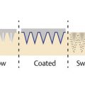
Similar to combining PRP with laser resurfacing, combination PRP plus microneedling has become an increasingly popular procedure. And there is a growing body of literature investigating the effects of microneedling with and without application of PRP for many indications. 29 A more in-depth discussion of these procedures can be found in Chapters 6 and 9. Briefly, it is likely that adding PRP to microneedling procedures aimed at skin rejuvenation improves outcomes and helps to minimize transient side effects as is seen when PRP is combined with laser resurfacing. 29 Sasaki used fluorescein-labeled platelets to attempt to further understand how PRP penetrates after microneedling procedures. 30 He used confocal laser microscopy to measure the absorption of PRP and determined that the optimal time for massaging PRP down 1.0 mm microneedling channels was between 5 to 30 minutes after microneedling. 30 Future studies need to done to further analyze how PRP penetrates the skin with application during both microneedling and laser resurfacing procedures.
3.5 Platelet-Rich Plasma Combined with Fat Grafting and Hyaluronic Acid Injections for Enhanced Augmentation
Combining PRP with other injectable substances such as autologous fat or hyaluronic acid filler is appealing, since the combination may provide synergistic effects. Augmentation with fat grafting and hyaluronic acid fillers offer impressive aesthetic results, but these treatments have risks of adverse reactions and/or lack longevity. 4 The addition of PRP may potentially decrease the volume needed for sustained correction, provide a scaffold for delayed biostimulation that increases longevity of temporary fillers, aid in engraftment of autologous fat, and help limit potential complications.
Ulusal published a series of 94 patients treated with intradermal injections of PRP combined with hyaluronic acid. 31 He found high patient satisfaction and a statistically significant improvement in skin firmness-sagging, skin texture, and general appearance based on ratings by three independent physicians as well as the patients themselves. 31 There were no significant complications reported. 31
Another study involved 31 patients treated with hyaluronic acid mixed with PRP delivered via injections and with a “skinroller.” 32 Subjects received 3 monthly treatments with outcomes assessed for 6 months after the final session. 32 When compared to baseline, patients demonstrated statistically significant improvement in skin firmness and elasticity and reported no serious complications. 32 Future controlled trials are necessary to compare the efficacy and safety of combination hyaluronic acid and PRP versus each product separately. It is imperative that these studies explicit outline the preparation protocols and final solution composition, as small changes in platelet, leukocyte, and erythrocyte concentration and dose may impact how the solutions interact and synergize. Application techniques, whether administered by injection or more superficially through microneedling pores, must also be examined rigorously.
The evidence for combining PRP with autologous fat grafting is controversial with some reports suggesting increased graft survival and hemostasis and others not. See Chapter 1 for a more in depth discussion. In short, Wellemsen et al reported a retrospective analysis of 82 patients with regard to recovery time and the aesthetic improvement treatment with fat grafting and minimal access cranial suspension (MACS)-lift with and without the addition of PRP. 33 Adding PRP to facial lipofilling reduced recovery time and improved the overall aesthetic outcome of a MACS-lift. 33 A randomized clinical trial by Fontdevila and colleagues found no difference in bilateral volume gain or maintenance via computed tomography imaging of grafts plus PRP versus grafts only, whereas two other blinded clinical trials looking different combination therapies did report benefits. 34, 35, 36, 37, 38 These differences may result from the diverse composition and preparation of PRP solutions. Inclusion of the buffy coat may have substantial impact on autologous fat graft survival due to inflammatory effects. So, like other PRP combinations, larger controlled trials with explicitly outlined, standardized techniques are still needed.
3.6 Techniques and Considerations
Although PRP has been utilized for over four decades in orthopedics, maxillofacial and other surgical fields of medicine, it has been adopted only more recently for aesthetic purposes, likely due to insufficient, small scale, or conflicting literature without clear objective evidence regarding its efficacy. 39, 40 Many systemic reviews have been conducted regarding the use of PRP for rejuvenation and augmentation. 39, 40, 41 In 2017, Frautschi et al published a comprehensive review searching for reports of PRP in aesthetic medicine published from 1950 to 2015. 39 After review of 38 reports, the authors concluded that the published studies produced promising, context-dependent results but lacked consistent reporting of preparation, composition, and activation methods, making any meaningful meta-analysis unrealistic. Moreover, the majority of studies were case series lacking controls and while most studies claimed effectiveness, objective measures were only used in 47% of the studies. 39 A review of studies on PRP in aesthetics from 2006 to 2015 by Motosko et al agreed that, although the majority of studies yield positive results and are safe, there is significant variation in preparation and treatment protocols, which makes conclusive findings difficult. 40 More rigorously designed studies for acne scarring, including randomized controlled and split face trials as well as flow cytometry reports, have been published since that time and lend further credence to this potential rejuvenate modality. 2, 42, 43, 44, 45
Because PRP is autologous, there is innate variability in PRP platelet concentration. 46 Platelet concentrations in humans can range from 150,000 to 450,000/μL, so this variation can potentially impact PRP concentration by threefold. 39 Most of the current studies do not account for baseline platelet levels or report the platelet concentration of PRP. There is also discussion whether concentration versus total absolute platelet dose/count may be better metrics to measure efficacy, since the latter is not volume dependent. 39 These studies also fail to account for the effect of various common pharmaceutical agents potentially affecting platelet function (i.e., aspirin, statins, antibiotics, and serotonin reuptake inhibitors), though a single small study suggests these medications may have little impact. 33
Another topic of much debate and perhaps equally important to platelet concentration/count is the inclusion or exclusion of other cell lines. Leukocytes and erythrocytes can substantially impact PRP’s activity and growth factor profile. Studies of high platelet-, high leukocyte-producing PRP systems report increased concentrations of many catabolic molecules. 47 White blood cells found in the buffy coat produce a large amounts of matrix metalloproteinases, 48 which may limit tissue repair and degrade extracellular matrix, as seen in photoaging. 49 Red blood cells release reactive oxygen species, alter solution pH, and may deposit hemosiderin, 47 all factor that can influence tissue response and resultant outcomes. Understanding the effects on PRP of these cell lines for each of the different aesthetic indications is critical to the adoption and standardization of such treatments. 47, 48, 49
The methods of PRP preparation remain variable and often poorly described, thus studies are difficult to interpret, replicate, or compare head-to-head. 50 PRP preparation methodology warrants increased attention. 38 In the authors’ opinion PRP preparation protocols must be better classified and standardized before expanding on the current literature and evaluating clinical outcomes. At a minimum, future studies should report clear preparation methods along with the starting and final cellular compositions of their therapeutic solutions. Frautschi et al recommended a classification system that can be used to help produce scientifically grounded conclusions and help facilitate a clearer understanding of situations in which PRP is most effective. 39 The proposed FIT PAW classification system includes seven components: (1) force of centrifugation, (2) sequence of centrifugation, (3) time of centrifugation, (4) platelet concentration, (5) anticoagulant use, (6) activator use, and (7) white blood cell composition. 39 The use of a reporting system such as this may help demystify PRP and allow for consistent results across different medical specialties and treatment sites, all prerequisites to the development of well-founded PRP applications in aesthetics. 39
Despite these limitations, reported methods of using PRP for rejuvenation and augmentation include: cutaneous injections, saturation with microneedling ( ▶ Fig. 3.1, ▶ Fig. 3.2, ▶ Fig. 3.3, ▶ Fig. 3.4) and laser resurfacing, as well as combination with fat grafting and injectable fillers. 34 PRP is commonly injected intradermally ( ▶ Fig. 3.5, ▶ Fig. 3.6, ▶ Fig. 3.7) and most cases in the literature report minimal complications with this method. There were reports of transient bruising and pain with periorbital intradermal injections of PRP. 26 One of the authors (J.B.) prefers to use a cannula for periorbital PRP injections ( ▶ Fig. 3.8) finding that this technique minimizes the incidence and severity of swelling and bruising, but acknowledging that it prohibits the plane of intradermal injection. PRP has been prepared with other active ingredients such as growth factor formulations and as discussed above, with autologous fat, and hyaluronic acid fillers. 17, 31, 39 The implications of such combinations are unclear, but the potential for synergistic outcome remains alluring. Further studies are needed to evaluate how combinations and delivery methods can be optimized to achieve the most effective outcomes.
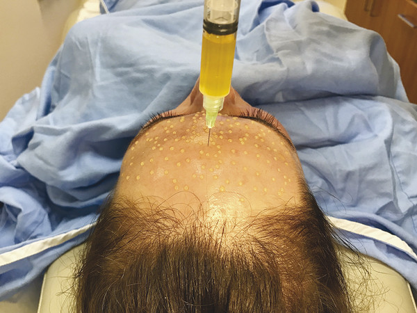
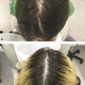
Fig. 3.1 PRP applied topically prior to microneedling.
Related posts:
 Platelet-Rich Plasma and Fibrin Sealants in Plastic Surgery: Clinical Applications and One Practice’s Experience
 Complications Associated with PRP and Microneedling in Aesthetic Medicine
 Microneedling: Mechanism and Practical Considerations
 Microneedling: Clinical Applications
 Applications and Safety in Skin of Color
 Combination Therapies
Stay updated, free articles. Join our Telegram channel

Full access? Get Clinical Tree


