Conditions treated
Nonscarring alopecia.
Actinic keratosis.
Dyschromia.
Rhytides.
Scars.
Striae.
Avoid
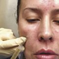
Active infection or inflammation (e.g., acne).
Prep
Mild cleanser.
Topical anesthetic.
Technique
Hyaluronic acid gel (or platelet-rich plasma in combination therapy) to facilitate device gliding.
Skin traction with perpendicular device tip placement.
Cross-hatch microneedling passes.
Pinpoint bleeding endpoint.
Ice water compression for hemostasis.
Postcare
Hydrating gel or cream (e.g., hyaluronic acid or hydrocortisone).
Mineral sunblock (SPF 30+).
6.1 Conditions Treated
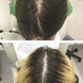
6.1.1 Microneedling for Alopecia
Microneedling (MN) has been shown to stimulate stem cells in the bulge region of the hair follicle, release growth factors through platelet activation and wound healing, and induce activation of important genes involved in phases of the hair growth cycle, including vascular endothelial growth factor (VEG-F), B catenin, Wnt3a and Wnt10b. 1 While the use of microneedling in alopecia is fairly limited, early studies show it to be of clinical utility.
Androgenetic Alopecia
Microneedling alone and in combination with topical minoxidil has been used in androgenetic alopecia (AGA) with positive results. A randomized, evaluator-blinded study compared the use of microneedling with 1.5 mm needles and twice-daily minoxidil 5% solution to the use of minoxidil alone in 100 men with mild to moderate androgenetic alopecia. Primary efficacy parameters were assessed at 12 weeks and included change from baseline hair count, patient assessment of hair growth, and investigator assessment of hair growth. The combination treatment group was statistically superior to the minoxidil only group in all three efficacy measures. 2
A subsequent case series of four male patients with stable AGA being treated with finasteride and minoxidil 5% solution received a series of 15 microneedling treatments over a 6-month period. Therapy with the finasteride and minoxidil was ongoing. Microneedling was performed on betadine-prepped scalp using a Dermaroller 1.5 mm with mild erythema as the clinical endpoint. After 8 to 10 sessions, new hair growth was observed in all four patients. Patient satisfaction ranged from 50 to 75% and results were sustained at 18 months follow-up. 3
A study comparing topical minoxidil to monthly sessions of combined platelet-rich plasma mesotherapy and scalp microneedling showed comparable results with regard to improvement of hair density; however, onset was significantly faster with minoxidil ( ▶ Fig. 6.1). 4

Fig. 6.1 Androgenic alopecia at baseline (left) and 3 months after third monthly microneedling session without concomitant topical therapy (right).
Alopecia Areata
Alopecia areata is an autoimmune inflammatory condition affecting hair follicles, which leads to usually discrete or less often diffuse patches of hair loss. Standard treatment with topical and intralesional corticosteroids is often ineffective or provides only temporary amelioration. Microneedling was shown to be effective in inducing hair growth in two patients with alopecia areata who had failed previous treatment with intralesional triamcinolone acetonide, topical steroids, and minoxidil 5% lotion. 5 Triamcinolone acetonide (10 mg/mL) was applied to the scalp both before and after microneedling using a Dermaroller with 1.5 mm needles. Three treatments were performed at 3-week intervals. Anesthesia was not necessary as the patients reported the procedure as painless. The investigators used pinpoint bleeding as their clinical end point and noted progressive improvement after each session. Three weeks following the final treatment, regrowth was considered excellent with results maintained 3 months posttreatment. The authors proposed that microneedling facilitated uniform and enhanced absorption of triamcinolone acetonide that could mitigate steroid-associated atrophy and limit the discomfort experienced with intralesional therapies.
6.1.2 Microneedling for Actinic Keratoses
Photodynamic therapy (PDT) is an approved effective treatment for treatment of actinic keratoses (AKs). Additionally, PDT has demonstrated cosmetic benefits. The effects of PDT are dose and time dependent and contingent, in part, on penetration of the photosensitizing agent. The stratum corneum (SC) is the main barrier to drug absorption. Physical pretreatment of the skin facilitates local uptake and is recommended to optimize results. 6 Studies showing increased protoporphyrin IX (PPIX) fluorescence suggest that microneedling prior to and following application of photosensitizers, such as methyl aminolevulinate (MAL) and delta amino levulinic acid (ALA), enhance their absorption. 7
Clementoni et al showed statistically significant improvement in global photoaging scores in 21 patients treated with combination red light and broadband pulsed light following microneedling at 0.3 mm depth than 1 hour incubation with 5-ALA. 8 However, lack of controls made it impossible to determine whether the clinical improvement was due to the combination of techniques or the PDT alone.
A split-face study compared conventional MAL–PDT preceded by curettage on one facial half to MAL–PDT followed by microneedling on the contralateral side. 9 After a 90-minute incubation, skin was irradiated with red LED. Side effects were more common, intense, and persisted longer on the side treated with a 1.5 mm microneedling device. No significant difference in actinic keratoses clearance rate between facial halves was noted, yet the microneedling-assisted PDT side had superior cosmetic results with greater improvement in mottled pigmentation, roughness, coarse wrinkles, fine lines and sallowness. The authors attributed the enhanced cosmetic improvement to better tissue delivery of the MAL and to the microneedle-induced wound healing response.
Another split-face study demonstrated both a greater decrease in mean percentage of actinic keratoses and increased cosmetic benefit on the side treated with microneedling prior to PDT compared to the standard PDT-treated side alone. 10 Microneedling was performed with a mechanical stamp style device at 0.5 mm. The skin was incubated with ALA for 1 hour prior to using the BLU U blue light for 1,000 seconds. It was unclear whether the cosmetic improvement resulted from the microneedling procedure itself, deeper penetration of the photosensitizer, or both.
A randomized study of 33 individuals with actinic keratoses revealed that 20 minute ALA incubation time following a microneedle roller (200 μm) with 1,000 seconds blue light exposure (total fluence 10 J/cm2) was superior to a 10 minute ALA incubation time. 11 Average actinic keratosis clearance was 76% with the 20 minute ALA incubation and 43% with the 10 minute ALA incubation, the latter not meeting statistical significance. The expedited 20-minute ALA incubation protocol mirrored the clinical efficacy typically achieved with conventional 1 hour ALA incubation time, thereby providing a suitable alternative to the time burden of standard PDT treatment.
While studies support the use of microneedling in conjunction with PDT, whether for treatment of actinic keratoses or improved cosmesis, it is important to note that other techniques can also facilitate absorption of photosensitizers. Bay et al found that the protoporphyrin IX accumulation was most enhanced following pretreatment with ablative fractional laser (10,600-nm fractional carbon dioxide [CO2]), followed by microdermabrasion, microneedling (0.2 mm Dermaroller) and curettage. 12 In clinical practice, however, practicality must be considered given that microneedling is minimally painful, significantly less expensive, faster to perform, and low downtime versus ablative fractionated laser treatment.
Improved efficacy of PDT has significant implications and may be particularly useful in organ transplant recipients. This population is immunosuppressed and has a substantially higher incidence of actinic keratoses compared to immunocompetent individuals. A study of 12 transplant recipients with actinic keratoses resistant to classical PDT showed a high clearance rate and low risk of recurrence after a series of three PDT treatments in which patients were pretreated with microneedling with a depth of 0.5 mm. 13
6.1.3 Microneedling for Dyspigmentation ( ▶ Fig. 6.2a, b)

Fig. 6.2 Melasma on the forehead at (a) baseline and (b) 6 months after one microneedling session and daily posttreatment use of mineral SPF 50 sunblock and topical vitamin C serum.
Melasma
Melasma is a common pigmentary disorder that is chronic and often recalcitrant to treatment. Photoprotection, topical treatments, chemical peels, and lasers have been used to treat melasma with varying results. Microneedling has been used with success to enhance melasma treatment results, although the mechanism of skin lightening is not yet well established. A study by Fabbrocini et al compared the use of microneedling (Dermaroller 0.5 mm) followed by application of a depigmenting serum (containing rucinol and sophora-alpha) on one side of the face with depigmenting serum alone on the contralateral side. Two treatments were performed at 1-month intervals. After each treatment, patients used a home roller device (Dermaroller-Model C8, 0.13 mm needle length) once-daily followed immediately by application of the depigmenting serum. Sunscreen was used on both sides of the face. Compared to the facial half treated with depigmenting serum alone, the combination therapy side had a statistically significant reduction in pigmentation and improved luminosity index. 14
A study of 22 patients with melasma recalcitrant to topical therapy and sunscreen were treated with two sessions of microneedling using a 1.5 mm Dermaroller at 1-month intervals. Twenty-four hours after the procedure, patients initiated daily application of a topical lightening agent (0.05% tretinoin + 4% hydroquinone + 1% fluocinolone acetonide) and SPF 60 sunscreen. The treatment was well-tolerated, and all patients responded and were satisfied with their clinical results. 15
Tranexamic acid (TA), a synthetic derivative of the amino acid lysine, is primarily used for its antihemorrhagic and antifibrinolytic properties in a surgical setting. 16 Topical TA has been found to decrease melanocyte tyrosinase activity through inhibition of ultraviolet (UV)-induced plasmin activity in keratinocytes. 17 Intradermal microinjections have been used as a treatment for melasma. 18 A study comparing microinjections of TA with topical TA both preceded and followed by at 1.5 mm microneedling (TA 4 mg/mL; Arm 1 with maximum 8 mg injected into a single area vs. Arm 2 with 4 to 5 cycles of 0.5 to 1 mL TA applied topically during needling) showed a better therapeutic response in the microneedling group, which was attributed to deeper and more uniform delivery of the TA. 19
Microneedling has also been shown to augment the response of melasma to laser treatment. A split-face study compared Q-switched (QS) Nd:YAG followed immediately by microneedling and vitamin C application to QS Nd:YAG alone for a series of four monthly treatments. 20 The combination treatment side demonstrated significantly better treatment response and greater improvement in MASI (Melasma Area Severity Index) scores. It was proposed that the QS Nd:YAG laser increased dermal circulation and thereby enhanced the microneedling mechanical effect and facilitated vitamin C penetration. It is important to note, however, that the risk of adverse events (particularly dermatitis) increases with the concomitant use of lasers and/or microneedling with topical products that are not intended for intradermal application.
Periorbital Melanosis
Microneedling has been used to successfully treat periorbital melanosis, a multifactorial condition often recalcitrant to treatment. Infraorbital dark circles were successfully treated in 13 women using a combination of microneedling followed by application of 10% trichloroacetic acid (TCA) for 5 minutes. Significant aesthetic improvement was seen in nearly all patients. 21
A case report using DermaFrac (Genesis Biosystems), which combines microneedling and simultaneous vacuum-assisted infusion of a serum, showed significant improvement of periorbital melanosis in an Indian male patient. 22 A total of 12 treatments were performed at 2-week intervals with application of either an “antiaging serum” or “lightening serum” (containing kojic acid). The favorable results reported may have been due to increased hydration and stimulation of new collagen and elastin reducing visibility of dermal pigment and blood vessels.
6.1.4 Microneedling for Skin Rejuvenation ( ▶ Fig. 6.3a, b)
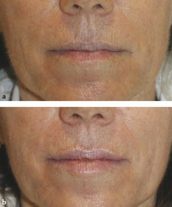
Fig. 6.3 Perioral rhytides at (a) baseline and (b) 3 months after one microneedling session.
Microneedling therapy has been successfully used as a minimally invasive treatment for skin aging. 23, 24 A study of 10 patients treated with six microneedling sessions at 2-week intervals showed histologic and clinical evidence of efficacy. 25 Biopsy specimens at 3 months confirmed epidermal acanthosis with rete ridge development. Interestingly, the total elastin content dropped, likely because solar elastotic material decreased while tropoelastin, a precursor to elastin, increased. This observation suggests that histology obtained at an early 3-month time point failed to capture the full tissue regeneration response and subsequent biopsies might have shown an overall rise in elastin content as the precursor tropoelastin aggregates into mature, well-organized rather than UV-irradiated fibers. New synthesis of collagen types I, III, and VII was also demonstrated. Results for collagen content were statistically significant at 3 months posttreatment and anticipated to improve further during the 1 year neocollagenesis process. 26 The collagen formed in the early phases of wound healing is type III then gradually replaced by collagen I, which remains in the area for 5 to 7 years. 23 Similar results were achieved in a retrospective analysis of 480 patients who received a series of one to four treatments. Increased collagen, in a normal lattice pattern, was observed 6 months posttreatment and a 40% increase in epidermal thickening was evident at 1 year. This study also found increase in elastic fibers at the later 6-month time points. 27
Microneedling has been utilized for hand rejuvenation, an increasingly popular area of concern. An initial feasibility study showed improved skin texture, skin tightening, and dermal neovascularization without dyspigmentation of dorsal hand skin after one microneedling treatment (depth unspecified). 28 Microneedling also successfully treated the aging neck in eight patients. Multiple modalities were used to assess efficacy including photographs, ultrasonographic images, and silicone rubber microrelief impressions. Two treatments led to a reduction in wrinkle severity grade in almost 90% of the patients. 29 Similarly, two sessions of microneedling of the upper lip showed marked reduction in wrinkle severity as assessed by photography and computer analysis of silicone rubber impressions of the wrinkles. 30
6.1.5 Microneedling for Scars
Numerous treatments exist for scar revision and often the best cosmetic outcomes are achieved by combining therapies. 31, 32
Acne Scars ( ▶ Fig. 6.4a, b)
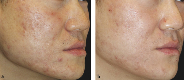
Fig. 6.4 Atrophic facial acne scars at (a) baseline and (b) 6 months after three microneedling sessions at monthly intervals.
Related posts:
 Platelet-Rich Plasma and Fibrin Sealants in Plastic Surgery: Clinical Applications and One Practice’s Experience
 Complications Associated with PRP and Microneedling in Aesthetic Medicine
 Microneedling: Mechanism and Practical Considerations
 Platelet-Rich Plasma for Rejuvenation and Augmentation
 Applications and Safety in Skin of Color
 Combination Therapies
Stay updated, free articles. Join our Telegram channel

Full access? Get Clinical Tree


