Department of Dermatology, Subharti Medical College, Meerut, Uttar Pradesh, India
Anup Kumar Tiwary
Keywords
JunctionalMelanocytic neviTheques
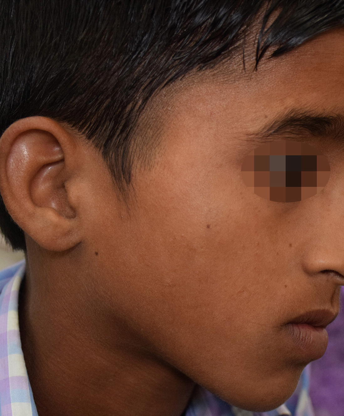
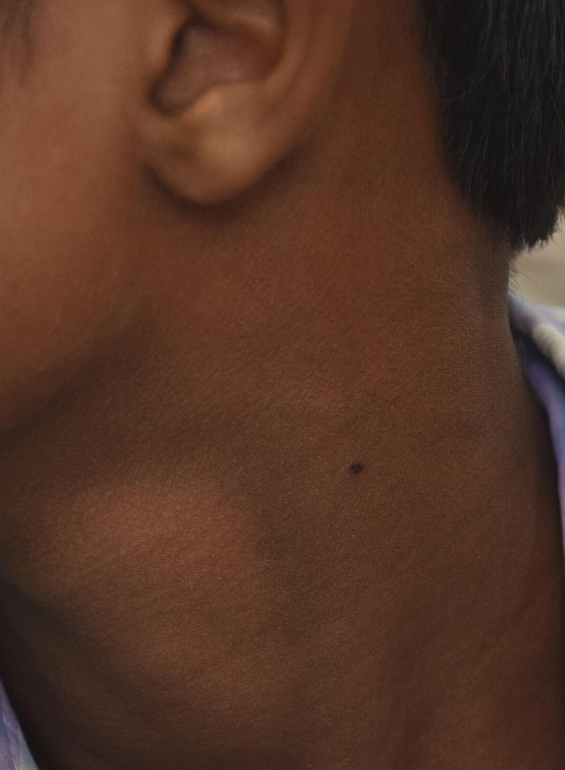
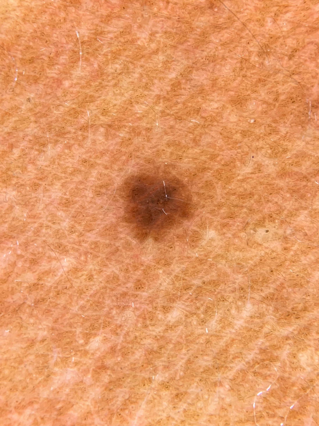
A 12-year-old boy came in dermatology department presenting with multiple, small, asymptomatic dark lesions on his right cheek and neck for past 2 years. The lesions appeared spontaneously without any history of trauma or dermatoses and have been static in size for past 1 year. The color remained same. No family history was present. On examination, there were three brown-black, well defined, macules of 3–4 mm size on right cheek (Fig. 23.1). Similar solitary brownish-black macule of about 4 × 3 mm size was noted on his neck on left side (Fig. 23.2). On dermoscopy, all lesions showed similar finding- multi-component pattern with peripheral reticular pigment network with blotchy pigmentationand pigmented globules in the centre (Fig. 23.3). Histopathology demonstrated nests of cuboidal melanocytes at the dermoepidermal junction, mainly located on the rete ridges.
Figure 23.1
Deeply pigmented small size macules on right cheek (Courtesy: Dr. Piyush Kumar)
Figure 23.2
Deeply pigmented small size macule on neck (Courtesy: Dr. Piyush Kumar)
Figure 23.3
Dermoscopic image showing multi-component pattern with peripheral reticular pigment network with central blotchy pigmentation and pigmented globules (Courtesy: Dr. Piyush Kumar)
What is the diagnosis?
1.
Congenital melanocytic nevus
Only gold members can continue reading. Log In or Register to continue