div class=”ChapterContextInformation”>
1. Bluish Gray Pigmented Macule on Right Cheek
Keywords
Dermal melanocytosisNevus of otaNevus fuscoceruleus ophthalmomaxillarisOculodermal melanocytosis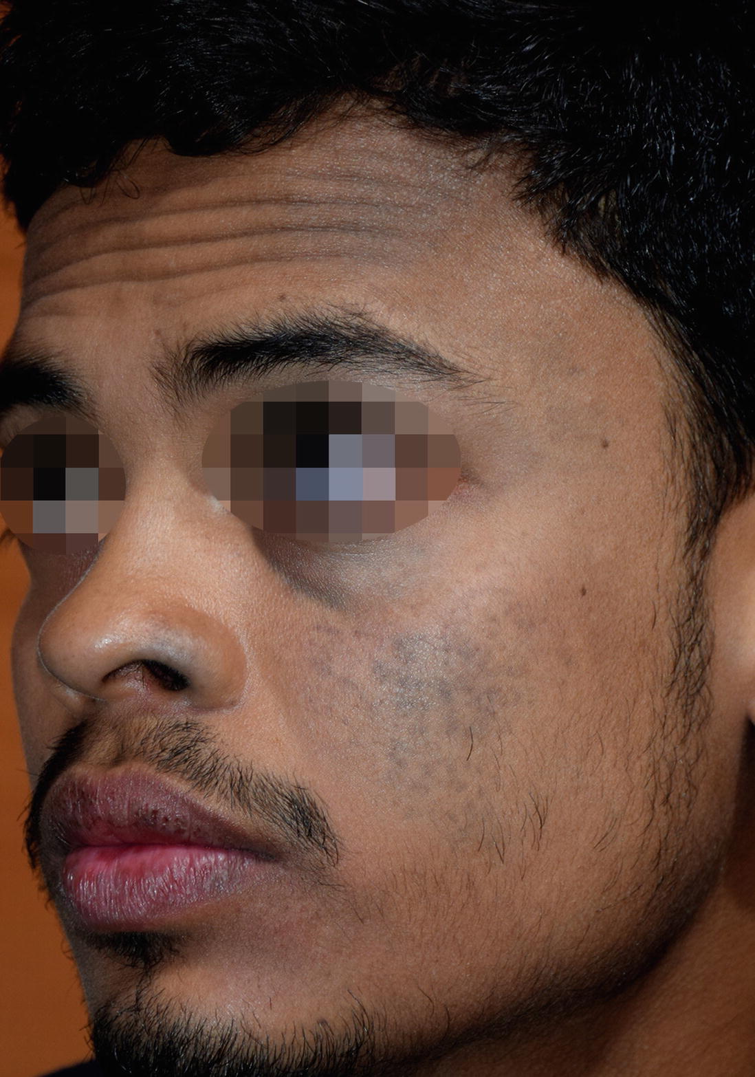
Young male is having bluish colored large macule on left cheek with scleral involvement
- (a)
Spilus nevus
- (b)
Nevus of Ota
- (c)
Café-au-lait macule
- (d)
Segmental lentigenosis
Histopathology showed elongated dendritic melanocytes around collagen bundles in superficial dermis.
Diagnosis
Nevus of Ota
Discussion
Nevus of Ota is a type of dermal melanocytosis characterized by unilateral bluish gray mottled pigmented macule with or without extracutaneous involvement. Histopathology shows dermal dendritic melanocytes deep within dermis. Various treatment modalities including lights and lasers had been tried with variable success.
During fetal development, normally melanocytes migrate from the neural crest to the dermal-epidermal junction (DEJ). However, these melanocytes may occasionally fail to reach at DEJ and remain entrapped in dermis where due to Tyndall effect brown color of these nevus cells give bluish gray color to skin surface. Exact etiology is not known but specific mutations have been detected within the dermal melanocytes, most often GNAQ or GNA11 suggesting a link between nevus of Ota and uveal melanoma. Several theories have been put forward which include: dropping-off of epidermal melanocytes, migration of hair bulb melanocytes, reactivation of pre-existing latent dermal melanocytes, which are triggered by dermal inflammation, UV radiation or hormonal changes during pregnancy [1].
Different types of dermal melanocytosis
Type | Epidemiology | Onset | Clinical | Distribution | Histopathology | Associated features |
|---|---|---|---|---|---|---|
Nevus of Ota | Asian (Japanese), female | Early infancy or adolescent | Blue-gray speckled pigmentation | In distribution of ophthalmic and maxillary branches of trigeminal nerve | Elongated dendritic dermal melanocytes in dermis | Glucoma Ocular melanoma Has been reported with Neurofibromatosis type 1 |
Nevus of Ito | Asian and female | Early infancy or adolescent | Blue-gray speckled pigmentation | In distribution of Acromioclavicular nerve | Elongated dendritic dermal melanocytes in dermis | None |
Mongolian spot | Asian, African and male | At birth or within first few weeks | Blue-gray uniform pigmentation | Lower back, sacral region | Spindle shaped dendritic melanocytes in deep dermis | Usually resolve within 1 year Extra sacral lesions tend to persist more than 1 year Persistent Mongolian spots are associated with inborn error of metabolism |
Nevus of hori | Asian and female | Second to fourth decade | Blue-gray speckled pigmentation | Area similar to nevus of ota but bilateral involvement | Elongated dendritic dermal melanocytes in dermis | None |
Dermal melanocytic hamartoma | Congenital | Blue-gray speckled macules in a diffuse pigmented patch | Dermatomal | Dermal melanocytes in upper second/third dermis | None |
Related posts:
 Years Old Male with Multiple Hyperpigmented Macules on Trunk
 Middle Aged Woman with Sudden Onset of Hyperpigmented Patch
 Female with Multiple Pigmented Macules on Face
 Young Female with Generalized Mottled Pigmentation
 Young Boy with Generalized Hyperpigmentation
 Female with Freckles Like Pigmentation on Face and Extremities
Stay updated, free articles. Join our Telegram channel

Full access? Get Clinical Tree


