div class=”ChapterContextInformation”>
3. Young Female with Multiple Pigmented Macules on Face
Keywords
LentigoFreckleUltraviolet exposureLentiginosisHyperpigmentation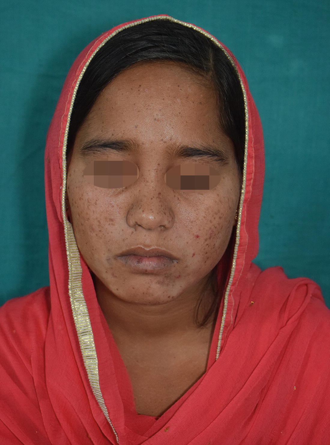
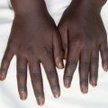
A young female with multiple discrete dark brown macules over face
- (a)
Freckles
- (b)
Simple lentigo
- (c)
Carney complex
- (d)
Centerofacial lentiginosis
Diagnosis
Simple lentigo
Discussion
Lentigines are hyperpigmented macules appearing on normal skin after ultraviolet (UV) rays exposure. There is increased number of melanocytes but not forming nests resulting in hyper pigmentation of basal layer which do not fade away after avoidance of UV exposure. Types of lentigines are: simple lentigo, solar lentigo, psoralene and UVA (PUVA) lentigo and ink-spot lentigo. Rarely, lentigines occur in association with hereditary multisystem syndromes.
Lentigines arise more commonly in light skinned individuals affecting children as well as adult in both sexes. Cause of lentigo depends on its type. Solar and ink-spot lentigo are associated with sun exposure, PUVA lentigo are associated with PUVA therapy and genetic factors are associated with other familial lentiginosis syndromes [1].
Lentigo simplex is the most common lentigo and chronic sun exposure is most important causing factor. Usually it starts in early childhood but sometimes lesions may present at birth or develop later. They gradually increase in number with age. Clinically lesions are round to oval, 3–5 mm in size, brown to black uniformly pigmented macules with jagged or smooth margin. They are asymptomatic and predominantly present over sun-exposure areas but may involve covered areas as well as mucosa without any other systemic involvement. Compared to freckles lentigines are darker in color and comparatively have sparseness in arrangement and scattered distribution. Avoidance of sun-exposure doesn’t lead to resolution of lesions [2].
Histopathology of lentigo simplex shows increased pigmentation of basal layer with slight increase in number of non-atypical melanocytes. Dermoscopy shows scalloped borders, pseudonetwork and structureless areas.
Lentigo simplex need to be differentiate from other types of lentigo, familial lentiginosis syndromes, ephelids (freckles), small junctional nevus, flat small seborrhoeic keratosis and lentigomaligna.
Types of lentigo
Related posts:
 Years Old Male with Multiple Hyperpigmented Macules on Trunk
 Gray Pigmented Macule on Right Cheek
 6 Years Old Male with Multiple Black Spots on Face
 Young Female with Generalized Mottled Pigmentation
 Young Boy with Generalized Hyperpigmentation
 Female with Freckles Like Pigmentation on Face and Extremities
Stay updated, free articles. Join our Telegram channel

Full access? Get Clinical Tree


