div class=”ChapterContextInformation”>
11. Itchy Pigmented Lesions on the Upper Back
Keywords
Primary localized cutaneous amyloidosisMacular amyloidosisAmyloid proteinHub and Spoke patternCongo redDimethylsulphoxide- 1.
Lichen planus pigmentosus
- 2.
Notalgia paresthetica
- 3.
Macular amyloidosis
- 4.
Poikiloderma of civatte
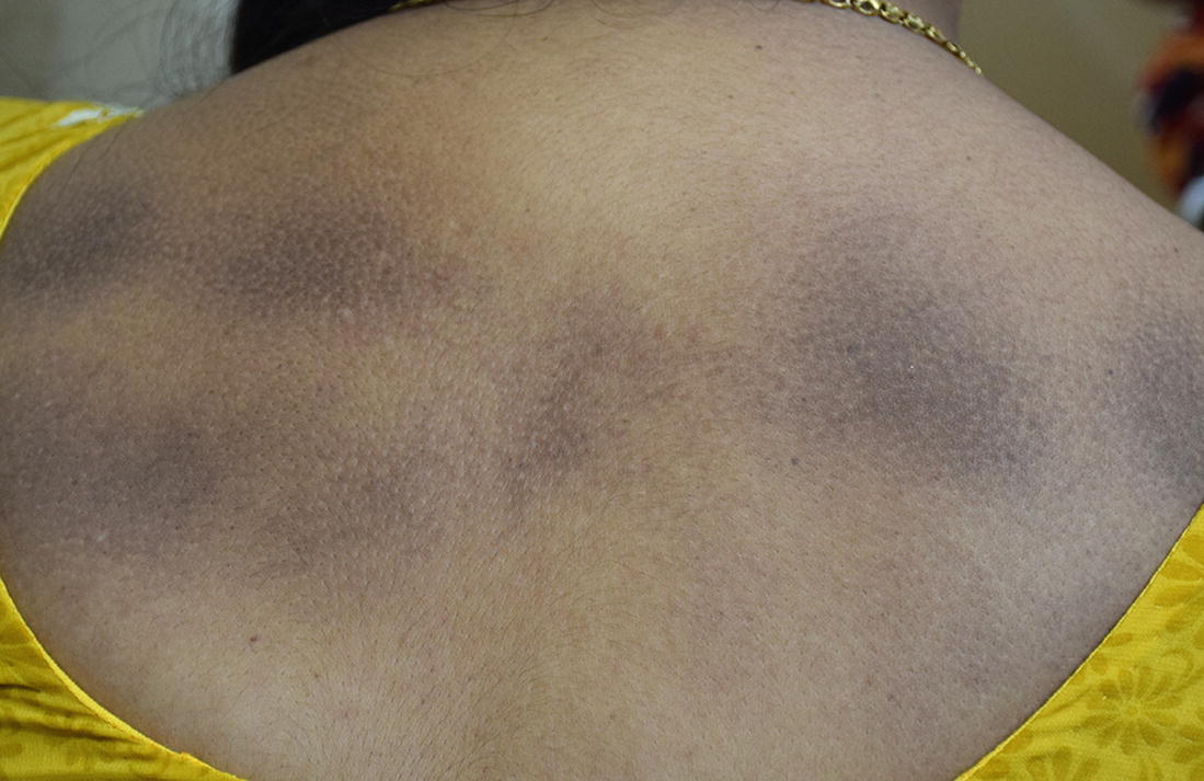
Multiple hyperpigmented patches on upper back
Multiple hyperpigmented macules arranged in a “rippled pattern”
Diagnosis
Macular amyloidosis
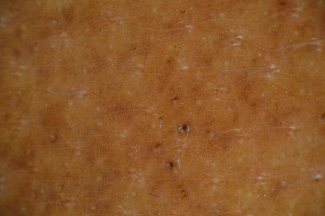
Dermoscopy of the back lesion showing brown clods with radiating brown lines, in a “Hub and Spoke” pattern (Courtesy: Dr. Shekhar Neema)
Discussion
The term “amyloid”, which stands for starch-like (Latin amylum) was introduced in science by Rudolph Virchow in 1854. The key feature of all types of amyloidosis is extracellular deposition of autologous proteins as characteristic amyloid fibril [1].
Macular amyloidosis (MA) represents a common variant of primary localized cutaneous amyloidosis (PLCA). The deposition of amyloid in previously apparently normal skin without deposits in the internal organs is known as primary localised cutaneous amyloidosis (PLCA). It is rare in western population, but has a high incidence in South-east Asia and some South American populations. Various subtypes of PLCA are recognized, including the more common macular and papular (lichen amyloidosis) types and the rare forms like the nodular variety and amylodosis dyschromia cutis. Both macular and papular lesions may occur in the same patient giving rise to biphasic amyloidosis [2]. Among all PLCA, only nodular amyloidosis has been found to be associated with systemic amyloidosis in 15–50% of patients and hence, long term follow-up of patients with nodular amyloidosis is recommended [3].
Multiple factors may play a collective role in the genesis of macular amyloidosis. These include racial, familial, and environmental factors, atopy, sunlight, friction, and female gender [4]. Amyloid deposits have also been shown to contain disulfide bonds, which are present in keratin. Based on this finding and on those of ultra-structural studies, cutaneous amyloid deposits are thought to be derived from degenerated keratin peptides of apoptotic keratinocytes transformed into amyloid fibrils by dermal macrophages and fibroblasts. A working hypothesis is that the epidermal trauma induced by long term scratching and rubbing results in keratinocyte degradation and formation of amyloid. Additionally, immunofluorescence studies with antikeratin antiserum have shown intense staining of the amyloid for the antikeratin antibody [5].
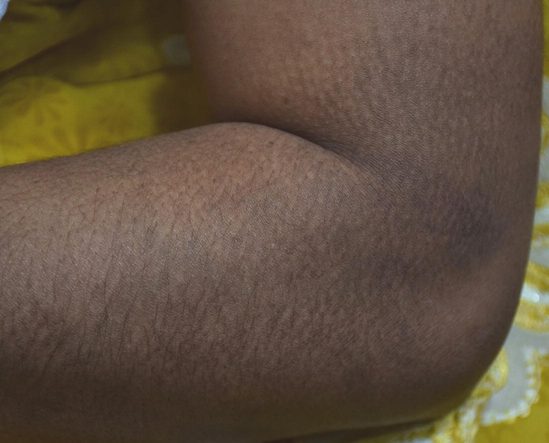
Clinically, MA presents as poorly delineated hyperpigmented patches consisting of grayish-brown macules, sometimes arranged in a “rippled pattern”. The most commonly involved sites are the interscapular area, and extensors of extremities (arms, shins and forearms), although involvement of the clavicles, breast, face, auricular concha, neck, and axilla have also been reported. It is usually pruritic (82%) and the degree of itching varies from mild to severe. It follows a chronic, progressive course with increasing pigmentation and extent of involvement. Other common PLCA is lichen amyloidosis which presents with intensely pruritic, discrete or coalescing, hyperkeratotic papules noted most commonly on the anterior tibiae. However, extensive lesions may involve extensor surfaces of the upper extremities and trunk. Amyloidosis cutis dyschromica is a rare type of PLCA characterized by the presence of both hyper- and hypopigmentation or depigmentation in reticular pattern distributed all over body [1].
Diagnosis of MA is made by its clinical presentation, dermoscopy, and histopathology. Histopathology with hematoxylin and eosin stain shows significant pigment incontinence with eosinophilic, amorphous deposits in papillary dermis which stain positive for amyloid with Congo red stain. Under polarized light, it gives green birefringence [5].
The common clinical differentials include lichen planus pigmentosus (LPP), notalgia paresthetica (NP) and poikiloderma of Civatte. LPP is clinically characterised by symmetrical distribution of dark brown to gray or gray-blue, round or oval macules with irregular and poorly-defined borders, which eventually enlarge and coalesce. It may be associated with variable itching and affects both sun-exposed and sun-protected areas of the body. Absence of rippled pattern helps in ruling out it as differential of macular amyloidosis. Histology reveals features similar to lichen planus with basal cell vacuolization and band like infiltrate at dermo-epidermal junction [6]. NP is a sensory neuropathy caused by entrapment of the posterior rami of spinal nerves T2 through T6 affecting the interscapular area and is associated with pain, paresthesia, hyperesthesia, or hypoesthesia rather than pruritus [7]. Poikiloderma of Civatte affects sun exposed areas in light skinned, perimenopausal females manifested by pink to brownish reticular patches consisting of linear telangiectasia, mottled hyperpigmentation and superficial atrophy. It affects the sides of neck, peripheral face and upper chest. Histology shows thinning of epidermis and solar elastosis in papillary dermis [8].
Treatment of MA is often disappointing. Topical treatment with corticosteroids with or without occlusion, 10% dimethylsulphoxide (DMSO), retinoids have been tried, but results are often unsatisfactory. Etretinate and acitretin therapy has been beneficial in some cases, but the condition seems to relapse after the treatment is stopped [9]. Dermabrasion may be beneficial on lichen amyloidosis of the shins. Oral cyclophosphamide and cyclosporine have a limited therapeutic efficacy [10]. In a study by Ostovari et al., 90% of patients with MA demonstrated more than 50% reduction in their pigmentation with the Q-switched Nd-YAG laser [11]. Ten and twenty percent trichloroacetic acid peels have also been tried with more than 50% improvement [9].
Key Points
Macular amyloidosis is a type of primary localised cutaneous amyloidosis in which amyloid deposit occurs in skin without involving internal organs.
Genetic and environmental factors result in focal epidermal damage with subsequent conversion of degenerated epidermal cell into amyloid in the papillary dermis which stain positive with Congo red.
Multiple small dusky brown-grey hyperpigmented macules arranged in rippled pattern is characteristic of macular amyloidosis.
Various treatment modalities have been tried with modest success.
Related posts:


Stay updated, free articles. Join our Telegram channel

Full access? Get Clinical Tree


