Fig. 13.1
A hemicircumareolar incision approach for patients with a large-diameter areola without breast ptosis
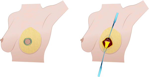
Fig. 13.2
A complete circumareolar incision associated with transdermic access along its inferior border for patients with a small/medium-diameter areola with some degree of breast ptosis
Relative contraindications include more significant breast ptosis (Regnault grade III or grade III [12]—Table 13.1), very large breasts, and especially a very small NAC (less than 2.0 cm). As the degree of ptosis increases, it is more likely that an L-shaped or inverted-T skin excision will be helpful in consistently achieving the desired result. If the nipple sits well below the inframammary fold or must be elevated more than 4 cm, a periareolar mastopexy becomes riskier. A very small NAC with a well-defined border is more likely to result in an enlarged areola and an unsatisfactory scar, with irreparable loss of the original shape and size of the areola.
Table 13.1
Regnault’s classification of ptosis
Degree | Characteristics |
|---|---|
Minor (grade I) | Nipple at the level of the inframammary fold |
Moderate (grade II) | Nipple below the inframammary fold, but above the lower breast contour |
Severe (grade III) | Nipple below the inframammary fold, at the lower breast contour |
Glandular ptosis | Nipple above the level of the inframammary fold but the breast hangs below the fold |
Pseudoptosis | Nipple above the level of the inframammary fold but the breast is hypoplastic and hangs below the fold |
13.3 Skin Markings
Usually, the skin markings (the sternum midline, the inframammary folds, and the areola diameters on the vertical lines from the midclavicle) are drawn with the patient in an upright position. If there is a large-diameter areola and no breast ptosis, a semicircular periareolar incision (hemicircumareolar) is usually indicated in BCS or nipple–areola sparing mastectomy (NSM). In these cases no additional skin markings are necessary. To prevent conspicuous scarring within or outside the areola, the incision is performed exactly at the junction of the areola and surrounding skin. If there is a small/medium-diameter areola and breast ptosis, an epidermic decortication of the complete circumareolar marked cutaneous ring and transdermic access along its inferior border are indicated (Figs. 13.2, 13.3). In these cases it is important to make the following marks: Point A, 19–21 cm from the midclavicular line and 10–12 cm from the external line. The ideal diameter of the NAC (25–30 mm) is outlined as a complete circumareolar epidermic ring (maximum width of 20–25 mm) which will be resected to reduce the cutaneous excess. The medial limit of resection coincides with the 10–12 cm of the external line and the same distance is maintained for the lateral limit. These limits of skin resection are confirmed by the medial–lateral and superior–inferior pinch test, ensuring there was no tension after removal of the skin.
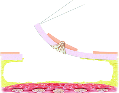

Fig. 13.3
The diameter of the nipple–areola complex (NAC) is outlined, as is the complete circumareolar epidermic ring, which will be resected. After the decortication, full access (transdermic) along its inferior border is performed. The transdermic access along the inferior border of the decorticated ring and glandular resection
13.4 Surgical Technique
The surgical procedure is performed with the patient under general anesthesia and with the patient’s the arms supported symmetrically 30° away from the chest. It is important to begin the sharp dissection with a no. 10 blade and the NAC is elevated off the underlying breast parenchyma Care is taken to leave a thickness of the retroareolar glandular tissue of approximately 1–2 cm to avoid nipple retraction. The incision is closed in layers with interrupted subcutaneous Vicryl 4-0 sutures and a continuous intracutaneous Prolene 4-0 suture (Ethicon, Johnson & Johnson, Hamburg, Germany).
In patients with a small/medium-diameter areola with breast ptosis, an epidermic decortication of the complete circumareolar marked cutaneous ring and transdermic access along its inferior border are performed. Thus, the subdermal plexus coming from the medial, lateral, and cephalic side of the areola is spared to ensure vascular supply to the NAC. Glandular excision is completed, leaving an adequate thickness of the subcutaneous tissue and a proper subareolar amount of gland. Skin flaps are handled carefully with the use of delicate hooks in order to maintain the integrity of the subdermal plexus and to avoid excessive skin flap traction. The skin is closed by the triple-layer technique. This technique is achieved by deepithelialization of the periareolar circle and advancement of the remaining areolar edge over this deepithelialized area. The advanced areolar edge is fixed over the deepithelialized area using a deeper layer of sutures anchoring the deepithelialized edge under the advanced areola and a superficial layer of sutures anchoring the edge of the advanced skin to the edge of the skin of the deepithelialized flap. This way, the terminal skin suture only overlies intact dermal and subcutaneous tissues, and all other sutures are not present in only one layer (Figs. 13.3, 13.4). A 2-0 nylon intradermal circumareolar purse-string suture is then used to limit the periareolar centrifugal tension and to improve areolar symmetry; a continuous 4-0 nylon suture (Ethicon, Johnson & Johnson, Hamburg, Germany) is used on the areolar skin surface to enhance the quality of the skin–areola transition zone.


Fig. 13.4
A partial submuscular pocket under the pectoralis major muscle is elevated from the inferior to the superior positions and the pectoralis muscle is partially detached. Closure of the periareolar incision is performed by the triple-layer technique. This technique is achieved by the advancement of the remaining areolar edge over this deepithelialized area. The advanced areolar edge is fixed over the deepithelialized area using a deeper layer of sutures anchoring the deepithelialized edge under the advanced areola and a superficial layer of sutures anchoring the edge of the advanced skin to the edge of the skin of the deepithelialized flap
13.5 Periareolar Technique in Skin-Sparing Mastectomy
SSM has been demonstrated to be an oncologically safe procedure for the treatment of early-stage breast cancer [1–3, 13, 14]. Compared with traditional mastectomy, SSM provides an ideal color and texture of breast skin and enhances the contour of the inframammary crease. To allow for adequate breast skin preservation, the oncoplastic surgeon should preoperatively discuss the periareolar incision and the width of the remaining skin flaps.
A critical survey of the literature shows that SSM is normally performed through numerous techniques, but most involve central breast incisions [1–3, 13, 14]. Habitually, the technique differs from surgeon to surgeon and is dependent on factors such as the type of reconstruction and the size of the breast. Although the type of incision differs, it is our impression that the best aesthetic outcome is related to the total periareolar approach [9]. With this technique, the final scar can be kept at the transition of the natural border of the future NAC (Fig. 13.5).
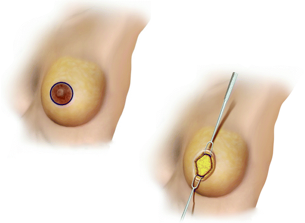

Fig. 13.5
The total periareolar approach for skin-sparing mastectomy (SSM) and immediate reconstruction. With this technique, the final scar can be kept at the transition of the natural border of the future NAC
In therapeutic SSM, specific areas of skin may in some instances require excision including prior incisions. Our approach to this issue is better performed through a central incision where the previous biopsy scar is excised in continuity with the NAC. Thus, previous communication of the team performing the biopsy/lumpectomy with the oncoplastic surgery team is critical in order to plan the incision as close as possible to the NAC. Usually, a total periareolar incision is performed, and with use of delicate hooks and fiber-optic retractors, the breast tissue dissection is performed in the same subcutaneous layer, reaching the final margins of the breast parenchyma. Skin flaps are handled carefully in order to maintain the integrity of the subdermal plexus and avoid excessive skin flap traction and even dermal exposure. A minimum flap thickness of 3–5 mm is maintained (Fig. 13.6).
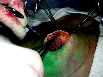

Fig. 13.6
The total periareolar incision is performed, and with use of delicate hooks and fiber-optic retractors, the breast tissue dissection is performed in the same subcutaneous layer, reaching the final margins of the breast parenchyma. Skin flaps are handled carefully in order to maintain the integrity of the subdermal plexus and to avoid excessive skin flap traction. A minimum flap thickness of 3–5 mm is maintained
The immediate reconstruction can be performed with an implant only, an expander and an implant, an implant associated with a pedicled latissimus dorsi muscle flap (LDMF), a transverse rectus abdominis musculocutaneous flap, or a deep inferior epigastric adipocutaneous free tissue transfer flap. In our experience, the reconstruction technique is frequently performed with a biodimensional implant–expander system associated with an LDMF as described elsewhere [9] (Figs. 13.7, 13.8). This option is usually chosen on the basis of individual aesthetic considerations but taking into account patient choice. This aspect is important since we have noted that some groups of patients prefer less evident breast scars. Thus, patients who are candidates for SSM and who do not want a large horizontal breast scar are the best candidates for SSM through a total periareolar incision. In fact, with the total periareolar incision and reconstruction, the final scar can be kept at the transition of the future NAC border, which may even be camouflaged by the NAC reconstruction. In addition, the latissimus dorsi muscle can be incorporated into the submuscular pocket. The implant–expander is placed in a total submuscular position, where a cover is made in its superior two-thirds by the pectoralis muscle and the inferior third by the LDMF. This allows creation of a tension-free muscular pocket while providing adequate tissue coverage for the implant–expander [9]. In cases in which vascularity of the mastectomy flap is unpredictable, the expansion can be initiated with limited fluid, which allows these flaps time to recover vascularity [9, 15, 16]. If there are small areas of skin necrosis, the patient can be treated on an outpatient basis with implant deflation and dressing changes since the implant is located under a healthy muscular pocket [9, 17]. In our experience, native breast skin complications were observed in almost 10 % of patients and represented one-third of all complications. Most cases consisted of partial skin loss and wound dehiscence between the LDMF and the breast skin [9].
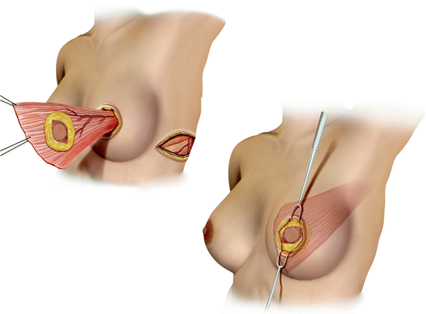
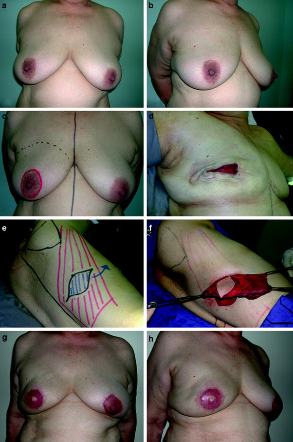

Fig. 13.7
The reconstruction technique is performed with a biodimensional implant–expander system associated with a latissimus dorsi myocutaneous flap. The flap provides adequate skin cover for the resected NAC and the final scar can be kept at the transition of the future NAC border. The latissimus dorsi muscle can be incorporated into the submuscular pocket and the implant–expander is placed in a total submuscular position where a cover is made in its superior two-thirds by the pectoralis muscle and the inferior third by the latissimus dorsi muscle flap

Fig. 13.8
A 54-year-old patient with a 4.8-cm invasive ductal carcinoma located in the right breast (a, b). The reconstruction markings showing the planned periareolar SSM (c). The patient underwent SSM with axillary dissection (d). The patient underwent immediate reconstruction with a biodimensional implant–expander (McGhan 150 volume 385–405 cm3) associated with a latissimus dorsi myocutaneous flap (e, f). Postoperative appearance at 11 months with a very good outcome after radiation therapy (g, h)
In spite of the main advantages, the total periareolar technique has some limitations. The surgical exposure is restricted and dissection can be troublesome if the oncological surgeon is inexperienced [1, 9, 13]. In this situation, more breast skin tension and flap irregularities can be noted and a poor exposure may result in an inadequate oncological resection (Fig. 13.8).


