Fig. 24.1
Oral mucosa involvement in pemphigus vulgaris. Erosions on the tongue
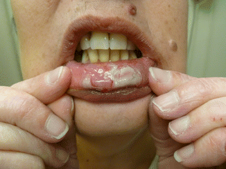
Fig. 24.2
Oral mucosa involvement in pemphigus vulgaris. Erosions on the lip
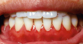
Fig. 24.3
Oral mucosa involvement in pemphigus vulgaris. Superficial, diffuse gingival erosions
Ocular involvement is rarely seen in pemphigus vulgaris and is frequently associated with a more severe course of disease. The most common ophthalmologic involvement is conjunctival but without progressive scarring such as occurs in ocular cicatricial pemphigoid [30]. Conjunctival involvement may occur in a unilateral or bilateral fashion: the presentation may range from mild hyperemia to conjunctivitis, resulting in symptoms of irritation and excessive watering of the eyes as well as the sensation of having a foreign body in the eye [31] (Fig. 24.4).
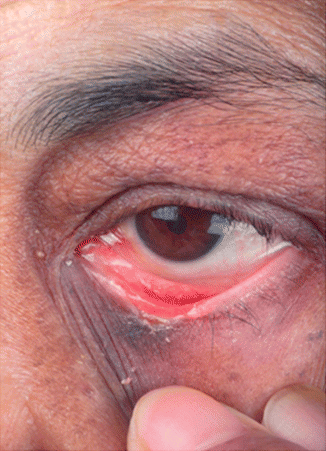
Fig. 24.4
Eye involvement in pemphigus vulgaris. Conjunctival hyperemia and conjunctival erosion involving lid margin on the left eye
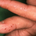
Genital involvement is less common than oral involvement and may be overlooked upon superficial clinical examination. Genital involvement usually occurs when there is extensive involvement of other sites. In most instances, the labia majora and minora are affected; involvement of the cervix is less common (Fig. 24.5). Pemphigus vulgaris localized to the vagina may present as chronic vaginal discharge and vaginal ulceration [32]. When the cervix is involved, some patients develop dyspareunia and cervical pemphigus may be misinterpreted as evidence of cervical dysplasia [33]. The mucosal surfaces that may be also involved with painful erosions include the nasal mucosa, penile skin, and anus [34–37].
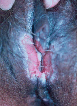
Fig. 24.5
Genital involvement in pemphigus vulgaris. Erosions on the labia majora and minora
24.3.2 Skin
Cutaneous involvement may be localized or generalized. Skin lesions can appear anywhere on the skin surface and arise on either normal-appearing skin or erythematous bases, but they have a predilection for the trunk, groin, axillae, scalp, face, and pressure points [38]. Flaccid blisters develop on these sites and may coalesce. The fluid within the bullae is initially clear but may become hemorrhagic or turbid. These blisters eventually rupture and result in erosions. Erosions have a tendency to spread at their periphery. This condition is characterized by Nikolsky’s sign; the direct Nikolsky is when the application of slight pressure on a blister results in extension of the blistering to adjacent skin and the indirect Nikolsky is when rubbing on clinically normal skin causes shearing. These signs are not always 100 % reliable for the diagnosis of pemphigus vulgaris, but they are suggestive if present [39]. The skin lesions in pemphigus vulgaris are rarely pruritic but are often painful [40]. The erosions soon become partially covered with crusts that have little tendency to heal (Figs. 24.6 and 24.7). Those lesions that do heal often leave hyper- or hypopigmentation with no scarring. Acanthomas may occur at sites of previous blisters [41]. Cases of cutaneous only pemphigus vulgaris are increasingly recognized [42]. In these cases, the antibody reactivity is still against Dsg3 and why the oral and other mucosae are spared is not yet understood.
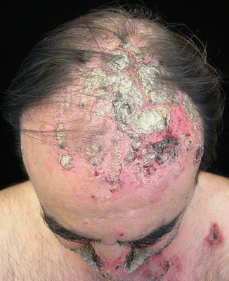
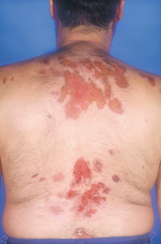
Fig. 24.6
Skin involvement in pemphigus vulgaris. Crusted erosions on the scalp
Fig. 24.7
Skin involvement in pemphigus vulgaris. Extensive erosions on the back
Involvement of the nails in pemphigus vulgaris is rare and is usually seen when the disease is severe [43]. The most common clinical manifestations include nail dystrophy, paronychia, and subungual hematomas [43, 44]. Fingernails are more frequently involved than toenails.
Pemphigus vegetans is a clinically distinct variant of pemphigus vulgaris which mainly affects large skinfolds such as the groins and axillae. Clinically, vegetating painful erosions are the major hallmark of this clinical variant (Figs. 24.8 and 24.9). Pemphigus herpetiformis is also considered as a clinical variant of pemphigus vulgaris which is characterized by vesicles and blisters which are grouped in a herpetiform pattern. Oral involvement is rather rare. Recent evidence suggests that pemphigus vegetans and pemphigus herpetiformis are associated with a particular autoantibody profile, i.e., the presence of IgG autoantibodies against desmocollin 3 [45]. The pathological role of these autoantibodies is not yet fully elucidated.
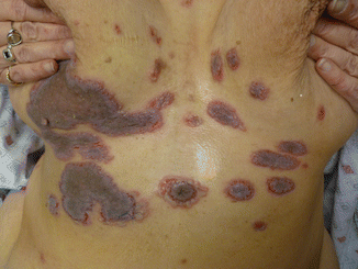

Fig. 24.8
Pemphigus vegetans. Vegetative erosions under the breasts
Fig. 24.9
Pemphigus vegetans. Vegetative erosions on the groins
24.4 Differential Diagnosis
There are a variety of blistering conditions that should be considered when seeing patients with pemphigus vulgaris including those of autoimmune, infectious, or inflammatory etiologies.
The differential diagnosis for mucosal lesions includes stomatitis secondary to herpes simplex virus, aphthous ulcers, lichen planus, paraneoplastic pemphigus, lupus erythematosus, Behcet’s disease, or dermatitis herpetiformis. The differential diagnosis for cutaneous involvement includes pemphigus foliaceus, pemphigus vegetans, IgA pemphigus, paraneoplastic pemphigus, bullous pemphigoid, linear IgA disease, erythema multiforme, candidosis, Crohn’s disease, Grover’s disease, and Hailey-Hailey disease.
24.5 Disease Activity Categorization
An international consensus statement on Disease Endpoint and Therapeutic Response for Pemphigus [46] divides pemphigus disease activity into the following stages of clinical evolution:
Early endpoints
Baseline
Control of disease activity
End of consolidation phase
Late endpoints
Complete remission off therapy
Complete remission on therapy
Minimal therapy
Minimal adjuvant therapy
Partial remission off therapy
Partial remission on minimal therapy
Relapse/flare
Treatment failure
Early endpoints provide a useful clinical indicator for clinicians regarding the commencement of differing treatment regimes. The baseline is classified as the day that the treating practitioner initiates treatment. Control of disease activity is defined as the time at which there is cessation of new lesions in conjunction with the healing of preexisting lesions. In the majority of cases, the expected time period in this stage is weeks. The end of the consolidation phase is the time period in which no new lesions have developed over a minimum period of 2 weeks. This phase is also characterized by the healing of most lesions, and most dermatologists would consider the weaning of steroids during this time period.
Late endpoints of disease activity may be reached with or without therapy. Complete remission off therapy is characterized by the absence of new lesions over a 2-month period after cessation of therapy. Minimal therapy constitutes treatment with less than or equal to 10 mg/day of prednisone or the equivalent or the use of minimal adjuvant therapy for a duration of at least 2 months. Minimal adjuvant therapy comprises of half the dose required to be defined as treatment failure. Partial remission off therapy is classified as development of lesions after cessation of treatment that heals within 1 week without treatment. Patients must be off systemic therapy for 2 months to be classified in this category. Patients may suffer a partial remission on minimal therapy when they develop new lesions that heal within 1 week while receiving minimal therapy. Topical steroids also constitute minimal therapy.
A relapse/flare is defined by the development of three or more new lesions that persist without healing for greater than 1 week or by the extension of preexisting established lesions. Treatment failure results when there is no change in disease activity despite treatment on therapeutic doses of systemic steroids and other agents whose doses and durations were agreed by international consensus [46].
References
1.
Lever WF. Pemphigus. Medicine (Baltimore). 1953;32(1):1–123.CrossRef
Related posts:
 Kindlin-1 and Its Role in Kindler Syndrome
 Cyclophosphamide in Autoimmune Blistering Diseases: Safety, Efficacy and Evidence Base
Management of Bullous Systemic Lupus Erythematosus
Cyclophosphamide in Autoimmune Blistering Diseases: Safety, Efficacy and Evidence Base
Management of Bullous Systemic Lupus Erythematosus
 Using Intravenous Immunoglobulins in Autoimmune Bullous Diseases
 Living with Epidermolysis Bullosa: Reviewing the Impact on Individuals’ Quality of Life
 Dermatitis Herpetiformis
Stay updated, free articles. Join our Telegram channel

Full access? Get Clinical Tree


