Host variables
Recipient age <1 or >10 years [10]
Prior damage to gut (viral illness, prolonged fasting) [12]
Donor or graft variables
HLA mismatch [2]
Use of unrelated donor [2]
ABO blood group mismatch [2]
Graft with high CD34+ cell dose or low regulatory T-cell content [12]
Other variables
Genetic polymorphisms within genes encoding for innate immunity, or inflammatory/immunoregulatory proteins in either donor or host [12]
Clinical Features of Acute GVHD
Although acute GVHD most often occurs within 1–2 months after HSCT [6, 15], the diagnosis can be made at any point after transplantation. Because time-based criteria are currently less emphasized, there is a greater emphasis on clinical features in making the diagnosis of acute GVHD [10, 15, 16].
Acute GVHD most commonly targets the skin, liver, and gastrointestinal tract [1, 17]. The skin is the most frequently affected organ and is often the first involved [15]. The classic rash is pruritic, may be painful, and is characterized by erythematous macules and papules coalescing on the trunk and extremities (often sparing the scalp), resembling the morbilliform rash of measles (Fig. 9.1). Acral involvement is common (Figs. 9.2 and 9.3). In severe GVHD, bullae and desquamation may develop, and with extensive involvement, may resemble toxic epidermal necrolysis (TEN) (Figs. 9.4 and 9.5). Gastrointestinal symptoms include nausea, vomiting, anorexia, abdominal pain, and diarrhea [15].
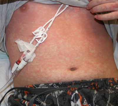
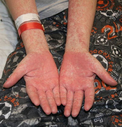
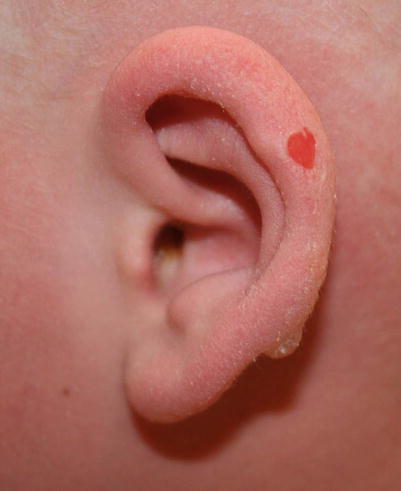
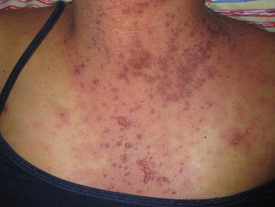
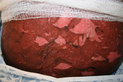
Fig. 9.1
Acute GVHD presenting as a morbilliform skin eruption
Fig. 9.2
Acral involvement in acute GVHD
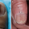
Fig. 9.3
Acral involvement (ear) in acute GVHD
Fig. 9.4
Bullous lesions are a poor prognostic sign in acute GVHD
Fig. 9.5
Toxic epidermal necrolysis–like acute GVHD
Differential Diagnosis for Acute GVHD
The differential diagnosis for acute GVHD includes bacterial/viral exanthem, engraftment syndrome, toxic erythema of chemotherapy, drug hypersensitivity reaction, and radiation recall dermatitis [1, 2].
Infectious exanthema occur more commonly in children [18] and solid-organ transplant and HSCT recipients are at increased risk for HHV6 and HHV7 reactivation, making infectious etiologies important to consider. Signs and symptoms of infection typically accompany the classic childhood exanthems, and the distribution and evolution of the rash may be helpful in differentiating these from acute GVHD. The viral exanthem of HHV6 is characterized by erythematous macules and papules surrounded by white halos, which begin on the trunk and spread to the neck and proximal extremities. It is accompanied by high fever (101–106 °F) and resolves over several days [18].
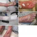
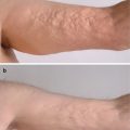
Engraftment syndrome occurs within days of granulocyte recovery and is characterized by fever >38.3 °C without a source of infection, rash over more than 25 % of body surface area that is not attributable to medication, and pulmonary edema [19]. Toxic erythema of chemotherapy (TEC) is a spectrum of cutaneous reactions to chemotherapeutic agents, most commonly presenting with erythema and tenderness of the palms, soles, and flexural regions including the axillae and groin (Fig. 9.6) [20]. There may be an increased incidence of TEC with conditioning regimens composed of busulfan and fludarabine, with a median onset of 22 days after dose administration [21].
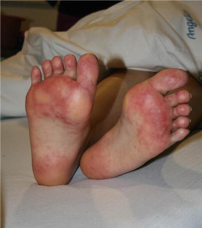
Fig. 9.6
Toxic erythema of chemotherapy presenting as tender, erythematous nodules on acral surfaces
Drug hypersensitivity reactions to non-chemotherapeutic agents should also be considered, although they tend to occur more in adults. Drug reactions typically occur within 1–14 days of initiating a drug, manifesting as a morbilliform rash on the trunk, which spreads to the extremities and less commonly involves the face, palms, or soles. Radiation recall dermatitis should also be considered in the setting of total body irradiation or recent history of sunburn followed by methotrexate for GVHD prophylaxis (Fig. 9.7).
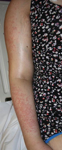
Fig. 9.7
Radiation recall dermatitis in a child with prior history of sunburn folowed by methotrexate for GVHD prophylaxis
Histopathology and Laboratory Evaluation of Acute GVHD
Though skin biopsies may confirm a diagnosis of acute GVHD if clinical suspicion is high, the histologic findings are nonspecific. Histologic findings include sparse lymphocytic interface and perivascular inflammation with variable degrees of adnexal extension. Dyskeratosis, spongiosis, lymphocytic exocytosis, and satellitosis may also be present. In addition to lymphocytic infiltration, eosinophils may be noted, making it difficult to distinguish acute GVHD from drug hypersensitivity reactions. In more severe acute GVHD, subepidermal clefting and full-thickness epidermal necrosis may be seen, mimicking toxic epidermal necrolysis [22, 23]. Many of the differential diagnoses show similar features, making biopsy unhelpful or misleading if wrongly interpreted [24–29]. Thus, clinical observation is key in making an accurate diagnosis, with close attention paid to time course, evolution of disease, and response to withdrawal of a potential offending agent.
Classification of Acute GVHD
Proper classification of acute GVHD is important, as this largely directs therapy. In 1974, Glucksberg devised the original staging system for acute GVHD, which was later modified during the Keystone Conference in 1994 [30]. The Keystone staging attempted to classify acute GVHD based upon the extent of skin, liver, and gut involvement, but the staging of pediatric gut GVHD was not discussed during the Keystone Conference, and stool output varies considerably between children and adults. The current proposal set forth by the University of Michigan and now utilized by the Mount Sinai Acute GVHD International Consortium redefines the Keystone criteria based on volume of diarrhea per kilogram of body weight, rather than absolute volume (Table 9.2) [32]. An additional consideration when staging pediatric acute GVHD is the difference in the distribution of body surface area between adults and children, as children have larger heads and smaller extremities than adults.
Table 9.2
Staging and grading of acute GVHD in children
Stage | Skin | Liver (bilirubin) | Upper GI | Lower GI (stool output per day) |
|---|---|---|---|---|
0 | No GVHD rash | <2 mg/dL | No or intermittent nausea, vomiting, or anorexia | <10 mL/kg/day or <4 episodes/day |
1 | Rash <25 % BSA | 2–3 mg/dL | Persistent nausea, vomiting, or anorexia | 10–19.9 mL/kg/day or 4–6 episodes/day |
2 | Rash 25–50 % BSA | 3.1–6 mg/dL | 20–30 mL/kg/day or 7–10 episodes/day | |
3 | Rash >50 % BSA | 6.1–15 mg/dL | >30 mL/kg/day or >10 episodes/day | |
4 | Generalized erythroderma + bullae | >15 mg/dL | Severe abdominal pain ± ileus or grossly bloody stool (regardless of stool volume) | |
Gradea | ||||
0 | None | None | None | None |
I | Stages 1–2 | None | None | None |
II | Stage 3 | Stage 1 | Stage 1 | Stage 1 |
III | Stage 0–3 | Stage 2–3 | Stage 0–1 | Stages 2–3 |
IV | Stage 4 or | Stage 4 or | Stage 0–1 | Stage 4 |
Treatment of Acute GVHD
Because of the difficulty in treating acute GVHD, there is a significant emphasis on prevention. Prophylactic regimens typically consist of one or a combination of the following agents: prednisone, cyclosporine, tacrolimus, sirolimus, methotrexate, mycophenolate mofetil, antithymocyte globulin, or alemtuzumab [2].
Once acute GVHD occurs, the prophylactic regimen can be adjusted and additional treatment may be considered, based on the grade of disease. Grade I acute GVHD, which is limited to the skin, usually has a favorable course and can be treated with topical corticosteroids and calcineurin inhibitors or narrow-band ultraviolet B phototherapy (nbUVB). For moderate to severe (grade II–IV) acute GVHD, high-dose systemic corticosteroids are employed as the first line of therapy [2, 33]. Unfortunately, only about half of patients are responsive to steroids [15, 32]. If there is no response to systemic corticosteroids after 2–7 days or if there is rapid progression within 48–72 h, second-line therapies should be considered. Table 9.3 outlines treatment agents subject to trials in pediatric patients.
Table 9.3
Treatment of moderate to severe (grade II–IV) acute GVHD in pediatric patients
Agent(s) | Study design and references | Patients, n (% pediatric) | Population with aGVHD, % | Treatment regimen | Response of aGVHD overall | Response of Skin aGVHD | Survival of patients with aGVHD |
|---|---|---|---|---|---|---|---|
First-line therapy | |||||||
Prednisone or MPred | Prospective, randomized, controlled, multi-institution trial [34] | 95 (34) | 100 | Low dose = 2 mg/kg/day IV MPred; High dose = 10 mg/kg/day IV MPred | At 5 days, 65 % of the low-dose treatment group responded and 71 % of the high-dose group responded | Not specified | 63 % at 3 years for both treatment groups |
Retrospective, single-institution chart review [35] | 443 (40) | 100 | 60 mg/m2 PO (or MPred IV equivalent, 48 mg/m2) for 14 days followed by an 8-week taper | At 28 days, 35 % achieved CR, 20 % achieved PR, 40 % achieved NR, and 5 % were NE | Not specified | 53 % at 1 year | |
Second-line therapies (steroid-refractory disease) | |||||||
Anti-thymocyte globulin (ATG) | Prospective, randomized, open-label, single-institution trial [36] | 100 (46) | 100 | ATG + steroids: 15 mg/kg equine ATG IV + 20 mg/m2 prednisone twice daily for 5 days followed by 8-week prednisone taper. Steroids: 20 mg/m2 prednisone PO or IV 3 times daily for 7 days followed by 8-week taper. All patients with skin aGVHD also received 0.1 % topical triamcinolone three times daily | No significant difference between groups. At 42 days, 76 % achieved response (CR or PR) and 24 % achieved NR in both treatment groups | No significant difference between groups. At 42 days, 79 % achieved response (CR or PR) in those treated with ATG + steroids | No significant difference between groups. 60 % at 100 days, 48 % at 6 months, and 40 % at 2 years in those treated with ATG + steroids |
Daclizumab | Retrospective, single-institution chart review [37] | 14 (100) | 71 | 2 mg/kg IV weekly for 8 weeks followed by 1 mg/kg IV weekly for 4 weeks | At 12 weeks, 45 % achieved CR, 18 % achieved PR, 27 % achieved NR, 9 % were NE | At 12 weeks, 50 % achieved CR and 20 % achieved PR | 50 % at the time of publication (follow-up time not specified) |
Extracorporeal photopheresis (ECP) | Prospective, nonrandomized, single-institution study [38] | 72 (100) | 100 | Twice weekly for 1 month, then every 2 weeks for 2 months, and then monthly for at least 3 more months, for a total of 22 procedures | At completion of treatment, 72 % achieved CR, 11 % achieved PR, and 17 % achieved NR | At completion of treatment, 78 % achieved CR, 13 % achieved PR, and 9 % achieved NR | 71 % at 5 years |
Prospective, nonrandomized, multi-institution study [39] | 77 (100) | 43 | Twice weekly for 1 month, then every 2 weeks for 2 months, and then monthly for at least 3 more months, for a total of 22 procedures | At completion of treatment (range 8–467 days), 54 % achieved CR, 21 % achieved PR, and 24 % achieved NR | At the completion of treatment (range 8–467 days), 76 % achieved CR | 69 % for responding patients vs. 12 % for non-responders at 5 years | |
Prospective, nonrandomized, single-institution study [40] | 73 (100) | 68 | 2–3 times weekly until clinical improvement, then twice weekly for 2 times, then twice weekly every other week for 3 times, and finally twice monthly | At the completion of treatment, 32 % achieved CR, 36 % achieved PR, and 32 % achieved NR | At the completion of treatment, 83 % achieved response (CR or PR) | 64 % at 1 year and 46 % at 5 years | |
Retrospective, nonrandomized, single-institution study [41] | 25 (100) | 60 | 2 consecutive days weekly for 1 month, then every 2 weeks for 2 months, and then monthly for 3 months, for a median total of 12 procedures (range 4–21) | At completion of treatment, 47 % achieved CR, 27 % achieved PR, and 27 % achieved NR | At completion of treatment, 80 % achieved CR | 100 % for responding patients vs. 15 % for nonresponders at a median of 1.6 years (range 0.8–4) | |
Retrospective, single-institution chart review [42] | 27 (100) | 78 | Twice weekly until clinical improvement for a median of 6 procedures (range 2–25) | At median of 167 days (range 4–1816), 52 % achieved CR, 38 % achieved PR, and the rest were NR | At median of 167 days (range 4–1816), 81 % achieved CR | 69 % at 1816 days for both acute and chronic GVHD | |
Etanercept | Prospective, randomized phase II, single-institution trial [43] | 160 (19) | 100 | Etanercept + steroids: 0.4 mg/kg etanercept SQ twice weekly for 8 weeks + 2 mg/kg/day IV MPred; Steroids: 2 mg/kg/day IV MPred | At 4 weeks, 69 % of etanercept + steroids group achieved CR vs. 33 % of the group treated with steroids alone | At 4 weeks, 81 % of the etanercept + steroids group achieved CR vs. 47 % of the group treated with steroids alone | No significant difference between groups: 69 % for those treated with etanercept + steroids vs. 55 % for those treated with steroids alone |
Infliximab | Retrospective, single-institution chart review [44] | 10 (100) | 100 | 10 mg/kg IV weekly for 3–4 doses | At a median of 11 days (range 5–17), 80 % achieved CR and 20 % achieved PR | Not specified | 40 % at 8–30 months |
Retrospective, multi-institution chart review [45] | 24 (100) | 75 | 10 mg/kg IV weekly for a median of 4 doses (range 1–58) | At 56 days, 69 % achieved CR | At 56 days, 86 % achieved CR | 67 % at 6 months; 13 % beyond 3 years | |
Mesenchymal stem cells (MSC) | Retrospective, multi-institution chart review [32] | 37 (100) | 100 | Median dose of 2 × 10⁶ cells/kg (range 0.9–3 × 10⁶ cells/kg) IV for a median of 2 infusions (range 1–13) | At completion of treatment, 65 % achieved CR, 22 % achieved PR, 14 % achieved NR or had progression | At median of 6 days (range 4–10), 57 % achieved CR, 23 % achieved PR, and 20 % achieved NR | 37 % OS at median follow-up of 2.9 years (range 1.7 months–6.7 years); 65 % for those who achieved CR vs. 0 % for those who did not |
Prospective, multi-institution pilot study [46] | 12 (100) | 100 | 2 × 10⁶ cells/kg IV twice weekly for 4 weeks | At completion of treatment, 58 % achieved CR, 17 % achieved PR, and 25 % achieved MR | At completion of treatment, 100 % achieved CR | 58 % at 100 days and 42 % at median follow-up of 611 days (range 427–1111) | |
Prospective, open-label, single-arm, multi-institution study [47] | 75 (100) | 100 | 2 × 10⁶ cells/kg IV twice weekly for 4 weeks | At 28 days, 61 % achieved response (CR or PR) and 39 % achieved NR (MR, stable, or progressive) | At 28 days, 44 % achieved CR, 32 % achieved PR, 12 % achieved NR, and 12 % were NE | 58 % OS at 100 days; 76 % for responders vs. 28 % for nonresponders | |
Mycophenolate mofetil (MMF) | Retrospective, single-institution chart review [48] | 14 (100) | 100 | Median initial dose of 40 mg/kg/day (range 30–74) PO was increased to median maximum dose of 60 mg/kg/day (range, 34–107) PO | At 8 weeks, 79 % achieved CR and 21 % were NE | At 8 weeks, 100 % achieved CR | 87 % at median follow-up of 35 months (range 14–86) |
Methotrexate (MTX) | Retrospective, single-institution chart review [49] | 35 (100) | 100 | 10 mg/m2 IV weekly | At 4 weeks, 37 % achieved CR and 9 % achieved PR | At 4 weeks, 52 % achieved CR, 17 % achieved PR, and 9 % progressed | 62 % at 6 months |
Retrospective, single-institution chart review [50] | 27 (100) | 37 | 3–10 mg/m2 IV or PO weekly for a median of 5 doses | At completion of treatment (1–7 weeks), 50 % achieved CR, 20 % achieved PR, 30 % were stable, and none progressed | At completion of treatment (1–7 weeks), 62 % achieved CR, 25 % achieved PR, 13 % were stable, and none progressed | 70 % at 6 months and 60 % beyond 14 months | |
Pentostatin | Prospective, single-institution, phase I trial [51] | 23 (22) | 100 | 1.5 mg/m2 IV for 3 days for 1–2 cycles | At 28 days, 64 % achieved CR, 14 % achieved PR, 9 % achieved MR, and 13 % progressed | At 28 % days, 81 % achieved CR, 6 % achieved PR, and 12 % achieved NR | 25 % at 1 year |
Outcomes of Acute GVHD
Though acute GVHD correlates with increased engraftment and graft-versus-tumor effect [5], unfortunately it does not correlate with survival in pediatric HSCT recipients. In fact, acute GVHD increases the risk of chronic GVHD by 11-fold, and steroid-refractory acute GVHD indicates a poor prognosis [5]. Mortality from acute GVHD ranges from 8 % for mild acute GVHD to 55 % for severe acute GVHD, and is usually due to infection, hepatic failure, or malnutrition [11, 12, 54]. Many of the aforementioned prophylactic and treatment strategies result in some response, but none have been shown to be more effective than the others. Furthermore, no therapies have been shown to decrease mortality or prevent progression to chronic GVHD [12]. As our understanding of the pathogenesis of acute GVHD expands, additional targeted therapies are likely to arise [55]. Until then, additional randomized controlled studies are needed to assess the safety and efficacy of these therapies in children.
Chronic Graft-Versus-Host Disease in Pediatric Patients
Introduction to Chronic GVHD
Chronic GVHD is a major cause of morbidity and mortality for children after HSCT. However, less is known about chronic GVHD in children than in adults because the incidence of HSCT is lower in children, and chronic GVHD occurs half as frequently in pediatric HSCT recipients. Most literature focuses on adult populations, with limited data regarding the spectrum of cutaneous disease, safety and efficacy of treatment, and outcomes in children.
Incidence of Chronic GVHD
In the United States, over 1500 allogeneic HSCTs are reported annually in patients less than 20 years old, and the incidence of chronic GVHD across all types of allogeneic HSCT is about 25 %, suggesting that at least 400 cases of chronic GVHD occur in children annually [56, 57]. This incidence is about one half that of adult populations.
The incidence of chronic GVHD varies widely by risk factors, from as few as 6 % of sibling umbilical cord-blood transplant recipients to 65 % of unmatched peripheral blood stem cell transplant recipients [8, 58, 59]. In cohorts that received HLA-matched bone marrow transplantation, approximately 27–35 % of children developed chronic GVHD, compared with 57–60 % of adults [60–62]. Males are 50 % more likely than females to be affected by chronic GVHD, in part because of the increased risk of chronic GVHD associated with female donor to male recipient transplantation [57].
Related posts:
 Grading and Treatment of Acute Graft-Versus-Host Disease
 Wound Care in the Management of Chronic Graft-Versus-Host Disease
 Clinical Presentation of Nonsclerotic Epidermal Chronic Graft-Versus-Host Disease and Hair and Nail Changes
 Dermal and Subcutaneous Chronic Graft-Versus-Host Disease
 Diagnosis, Staging, and Treatment of Chronic Graft-Versus-Host Disease
 Clinical Presentation of Nonsclerotic Epidermal Chronic Graft-Versus-Host Disease and Hair and Nail Changes
Stay updated, free articles. Join our Telegram channel

Full access? Get Clinical Tree


