Pectoralis Flap for Chest Wall Reconstruction
Jeff J. Kim
David H. Song
DEFINITION
Chest wall defect: Most common etiologies include trauma, tumor resection, osteoradionecrosis, deep sternal wound infections, and chronic empyemas.
Chest wall functions: Protection of visceral organs, support of respiratory mechanics, and base scaffold for shoulder/upper limb
Basic defect management concepts: Adequate debridement of infection/nonviable tissue, reconstruct to re-establish form and function, replacing like with like whenever possible
Reconstructive goals:
Stabilize thoracic skeletal defect to return proper respiratory mechanics.
Obliterate intrathoracic dead space that may predispose to infection.
Protect vital intrathoracic structures.
Provide soft tissue coverage for closure.
Recreate aesthetic contour.
Defects requiring skeletal support to prevent paradoxical motion: 5 cm or more in diameter or two or more rib resection anteriorly, twice as much for lateral and posterior defect; radiated tissue can often tolerate larger defect due to rigidity from fibrosis.
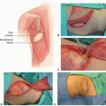
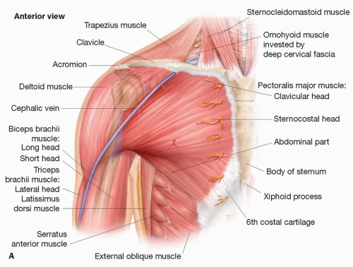 FIG 1 • A. Muscular anatomy of anterior chest and shoulder. |
ANATOMY
Pectoralis major
Origin: Sternum, 1st to 6th costal cartilage, clavicle (FIG 1A,B)
Insertion: Lateral lip of bicipital groove of humerus
Innervation: Medial and lateral pectoral nerve from medial, lateral cord of brachial plexus
Function: Adduction, extension, and medial rotation of shoulder
Borders: Superficial to pectoralis minor, superior and superficial to serratus anterior, inferior to the subclavius; two heads converge laterally inferior to deltoids; lateral border forms the anterior axillary fold/wall
Vascular anatomy (FIG 1C)
Mathes and Nahai classification: type V muscular flap—thoracoacromial artery (dominant) and internal mammary perforators (secondary segmental)
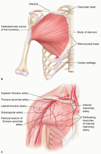
FIG 1 (Continued) • B. Origin and insertions of the pectoralis major. C. Vascular anatomy of blood supply to pectoralis major.
Thoracoacromial artery arises from second portion of axillary artery deep to pectoralis minor, travels laterally and merges superomedially to pectoralis minor, and divides into four branches as it pierces the clavipectoral fascia. The dominant branch to the sternocostal portion is the pectoral branch, which courses deep to pectoralis major after piercing through the clavicopectoral fascia medial to pectoralis minor before dividing into muscular and cutaneous branches; clavicular branch arising lateral to pectoralis minor has much shorter pedicle (1 cm) before exclusively supplying the clavicular head, subclavius muscle, and soft tissue around the clavicle.
Internal mammary artery (IMA) provides reliable secondary segmental pedicles through its perforating branches that pass through the intercostal spaces 1 to 2 cm lateral to the sternal edge; usually three perforators provide the muscle through the first to third intercostal space with branches coming through first or second space being the most dominant.
Venous drainage: Muscle drains mainly via venae comitantes of the pedicles, through pectoralis branch into the axillary vein, while overlying skin drains through venules accompanying arterial perforators.
PATHOGENESIS
Most common etiology and mechanism of chest wall defects requiring reconstructive interventions include injury from trauma, iatrogenic (radiation, cardiothoracic surgery), infection, congenital deformity, and neoplasm.
NATURAL HISTORY
Tansini is often credited with first use of muscle flap in chest wall reconstruction using latissimus dorsi.1
Jurkiewicz and others transposed the pectoralis major into the mediastinum either based on the thoracoacromial pedicle or as a turnover flap based on perforators of the IMA.2
Nahai introduced a modification of the turnover pectoralis major flap by dividing the muscle medial to the thoracoacromial pedicle and using only the medial two-thirds of the pectoralis major for definitive coverage of the mediastinal wound.3
Tobin suggested the splitting of the pectoralis major muscle into sternocostal, external, and clavicular segments and the preserving of the thoracoacromial pedicle.4
Morain suggested the splitting of the pectoralis major muscle into segments but leaving some of the segments intact to preserve muscle function for smaller defects requiring only a small portion of muscle.5
PATIENT HISTORY AND PHYSICAL FINDINGS
Preoperative evaluation for patients requiring pectoralis flap for chest wall reconstruction is mostly based on history and physical examination.
History
Etiology, chronicity
Important to note history of radiation and history of surgery, including history of coronary artery bypass grafting (CABG)/IMA harvest and other chest incisions. Also important to note pulmonary function and history of chronic obstructive pulmonary disease
Social: Smoking history
Physical examination
Important to note size, location, and tissue composition of defect
Soft tissue: Size and location
Skeletal: Number of ribs and size of skeletal defect
Intrathoracic: Size and volume of dead space
Respiratory mechanics: Skeletal stability, presence of paradoxical motion, soft tissue compliance
Previous scars, radiation changes
IMAGING
Preoperative computed tomography (CT) is not absolutely necessarily from a reconstructive standpoint, especially in wounds requiring further debridement where final size and composition of defect will invariably change.
If a CT scan is available, however, it can provide some preoperative insight for operative planning in some cases.
DIFFERENTIAL DIAGNOSIS
Trauma
Empyema
Poststernotomy mediastinitis
Osteoradionecrosis
Tumor resection of anterior chest wall
NONOPERATIVE MANAGEMENT
For small and superficial wounds or defects with no exposed vital structure or functional defect, it may be possible and appropriate to manage using simple wound care methods with either traditional wet to dry gauze dressings or negative pressure wound therapy.
In cases of heavily contaminated or actively infected wounds (except deep sternal infections), local wound care may be more appropriate initial method of management to prepare the wound bed prior to proceeding with definitive operative reconstruction.
SURGICAL MANAGEMENT
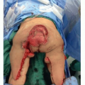
Pectoralis major offers large muscular flap with robust blood supply, with arc of rotation based on thoracoacromial pedicle by allowing coverage of central, supraclavicular, and axillary or lateral chest wall defects, as well as being able to be transposed to an intrathoracic position for obliteration of dead space.
The pectoralis major is most commonly used as an advancement flap to cover anterior chest wall defects in the upper part of the chest, classically for wounds resulting from an infected median sternotomy after open heart surgery.6
This technique allows for repeat subsequent procedures through the midline incision, including further debridement and costochondral resection, without sacrificing viability of reconstruction.1
Turnover flap is based off its medial segmental blood supply from perforators of IMA allows coverage of wounds extending further inferiorly or to the contralateral side or to provide large bulk of healthy tissue to obliterate more significant area of dead space.Related posts:

Stay updated, free articles. Join our Telegram channel

Full access? Get Clinical Tree