5
Paronychia
Sam Moghtaderi and Kevin D. Plancher
History and Clinical Presentation
A 34-year-old, right hand dominant woman presented to her physician complaining of a red, tender area surrounding the nail of the third finger of her right hand. She had received a manicure 3 days prior to the onset of symptoms.
Physical Examination
The periungual areas of the affected digit appeared erythematous and swollen, and were tender to palpation over the radial paronychial fold and the eponychium (Fig. 5–1). There was also elevation of the proximal nail bed with pus extruding from below.
Diagnostic Studies
The diagnosis of paronychia is generally clinical, and does not require any diagnostic studies.
PITFALLS
- Must differentiate from herpetic infections, for which incision and drainage (I&D) is typically contraindicated
- In making incisions, care must be taken to avoid damage to nail matrix
- Nonsurgical treatments typically not effective for chronic paronychia
PEARLS
- Paronychia is the most commonly seen infection of the hand
- Warm soaks and antibiotics effective if there is no drainage or fluctuance
- Eponychial marsupialization for chronic paronychia; also remove nail if signs of involvement
Differential Diagnosis
Herpetic whitlow
Acute paronychia
Chronic paronychia
Diagnosis
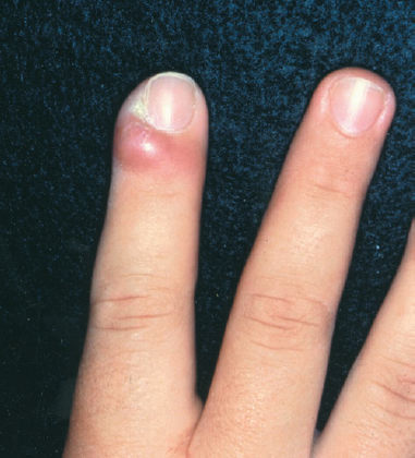
This patient’s presentation is typical of acute paronychia, an infection of the soft tissue folds surrounding the fingernail. The clinical presentation initially consists of localized tenderness of the paronychial region, with subsequent erythema, swelling, and fluctuance (Fig. 5–2). Frank drainage and elevation of the nail plate are also seen.

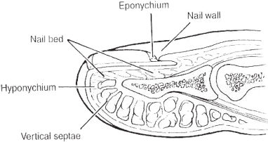
Figure 5–1. Schematic anatomic drawing of nail complex and its components.

Figure 5–2. Acute paronychia, with characteristic erythema and swelling.
Stay updated, free articles. Join our Telegram channel

Full access? Get Clinical Tree


