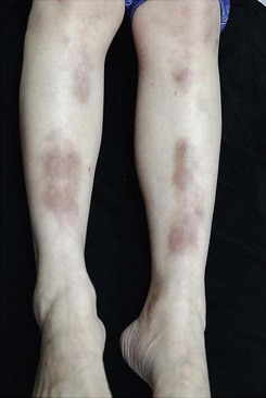
158 Necrobiosis lipoidica Arif M. Aslam and Ian Coulson Evidence Levels: A Double-blind study B Clinical trial ≥ 20 subjects C Clinical trial < 20 subjects D Series ≥ 5 subjects E Anecdotal case reports (From Pei-Shan Yen, Kuo-Hsien Wang, Wei-Yu Chen, Ya-Wen Yang, Wen-Tsao Ho, 2011. The many faces of necrobiosis lipoidica: a report of three cases with histologic variations. Dermatologica Sinica 29 (2), 67–71.) Necrobiosis lipoidica (NL) is a chronic cutaneous granulomatous condition with degenerative connective tissue changes. The pathophysiology remains unknown but both granulomatous and angiopathic mechanisms have been proposed. It is seen in 1 in 300 diabetics but may be unassociated with glucose intolerance. NL initially appears as an atrophic plaque found over pretibial sites. Ulceration may occur after trauma, with ensuing pain. NL may rarely be complicated by squamous cell carcinoma. Management strategy Smoking cessation and avoiding trauma to the affected shins are key factors to avoid transformation from an unsightly plaque into a painful, recalcitrant ulcer. The progression of new lesions may be halted by intralesional or occluded potent topical corticosteroids applied to the margins of the lesions. Once atrophy has developed there is little that will reverse this, although topical retinoids may be tried. Telangiectasia is often marked and has been treated with pulsed dye laser. Extensive lesions may justify trials of nicotinamide or prednisolone. Antiplatelet therapy in the form of aspirin, dipyridamole, or ticlopidine has its enthusiasts, though responses are inconclusive. Topical psoralen and UVA (PUVA) has received recent interest and may arrest progression and improve the appearance. A variety of systemic anti-inflammatory and immunosuppressive agents have received recent attention, including mycophenolate mofetil, fumaric acid esters, cyclosporine, antimalarials, thalidomide, and pentoxifylline. Infliximab and etanercept have also been proposed. The chronically ulcerated lesion is a challenge; antibiotics deal with secondary infection, appropriate dressings may be required, and growth factors such as becaplermin and granulocyte–macrophage colony-stimulating factor (GM-CSF) may accelerate healing. As diabetics may have coexisting large vessel atherosclerosis that may contribute to ulceration, non-invasive arterial studies or angiography need to be considered if clinically indicated. Venous hypertension may also contribute to the localization and ulceration of necrobiosis. Excision and grafting may transform the patient’s quality of life and improve cosmesis. Work with the diabetes specialist to optimize diabetic control. Specific investigations Two-hour postprandial glucose Skin biopsy Consider angiography or venous circulation studies Consider a biopsy to exclude sarcoidosis, which may mimic necrobiosis lipoidica, or, if clinical features suggest, the rare development of squamous cell carcinoma Dermoscopy Squamous cell carcinoma arising in an area of long-standing necrobiosis lipoidica. Lim C, Tschuchnigg M, Lim J. J Cutan Pathol 2006; 33: 581–3. Case report of a squamous cell carcinoma arising de novo in an area of NL. Carcinoma cuniculatum arising in necrobiosis lipoidica. Porneuf M, Monpoint S, Barnéon G, Alirezai M, Guillot B, Guilhou JJ. Ann Dermatol Venereol 1991; 118: 461–4. Although rare, squamous cell carcinomas may complicate any condition in which papillary dermal scarring is appreciable. Unilateral necrobiosis lipoidica of the ischemic limb – a case report. Naschitz JE, Fields M, Isseroff H, Wolffson V, Yeshurun D. Angiology 2003; 54: 239–42. A possible ischemic pathogenesis of NL emerges from a case of unilateral large vessel arteriosclerotic ischemia with ipsilateral NL. The author has experience of a severely ulcerated area of NL that only started to heal after the disobliteration of severe femoropopliteal atheroma. Dermatoscopy of early onset necrobiosis lipoidica. Bakos RM, Cartell A, Bakos L. J Am Acad Dermatol 2012; 66: e143–4. Dermatoscopy may be a useful tool for evaluating early onset NL before biopsy. The presence of branching telangiectasia, hairpin-like vessels and a yellow background may suggest this diagnosis. First-line therapies Stop smoking and optimize diabetic control C Intralesional or topical corticosteroids under occlusion D Necrobiosis lipoidica diabeticorum: association with background retinopathy, smoking, and proteinuria. A case controlled study. Kelly WF, Nicholas J, Adams J, Mahmood R. Diabet Med 1993; 10: 725–8. The patient should stop smoking and control diabetes mellitus with vigilance. Fifteen diabetic patients with NL were each matched with five control subjects with diabetes mellitus. Background retinopathy, proteinuria, and smoking were all more common with NL. No differences were noted between those with NL and controls in the prevalence of vascular disease and neuropathy. Glycosylated hemoglobin concentrations were higher in patients with NL. Granuloma annulare and necrobiosis lipoidica treated by jet injector. Sparrow G, Abell E. Br J Dermatol 1975; 93: 85–9. Three of five cases of NL underwent complete resolution and one had partial improvement with 5 mg/mL triamcinolone injection to the edges of lesions. No serious complications of this type of treatment were observed. Treatment of psoriasis and other dermatoses with a single application of a corticosteroid left under a hydrocolloid occlusive dressing for a week. Juhlin L. Acta Derm Venereol 1989; 69: 355–7. A 0.1% betamethasone alcoholic lotion under a hydrocolloid dressing was an effective, well-tolerated treatment, and three applications only were required. Second-line therapies Systemic corticosteroids D Aspirin and dipyridamole C Ticlopidine D Nicotinamide D Clofazimine D Topical PUVA D Topical tacrolimus E Only gold members can continue reading. Log In or Register to continue Related Related posts: Cat scratch disease Hemangiomas Drug eruptions Erythropoietic protoporphyria Ichthyoses Jellyfish stings Stay updated, free articles. Join our Telegram channel Join Tags: Treatment of Skin Disease Comprehensive Therapeutic Strategies Aug 7, 2016 | Posted by admin in Dermatology | Comments Off on Necrobiosis lipoidica Full access? Get Clinical Tree
158 Necrobiosis lipoidica Arif M. Aslam and Ian Coulson Evidence Levels: A Double-blind study B Clinical trial ≥ 20 subjects C Clinical trial < 20 subjects D Series ≥ 5 subjects E Anecdotal case reports (From Pei-Shan Yen, Kuo-Hsien Wang, Wei-Yu Chen, Ya-Wen Yang, Wen-Tsao Ho, 2011. The many faces of necrobiosis lipoidica: a report of three cases with histologic variations. Dermatologica Sinica 29 (2), 67–71.) Necrobiosis lipoidica (NL) is a chronic cutaneous granulomatous condition with degenerative connective tissue changes. The pathophysiology remains unknown but both granulomatous and angiopathic mechanisms have been proposed. It is seen in 1 in 300 diabetics but may be unassociated with glucose intolerance. NL initially appears as an atrophic plaque found over pretibial sites. Ulceration may occur after trauma, with ensuing pain. NL may rarely be complicated by squamous cell carcinoma. Management strategy Smoking cessation and avoiding trauma to the affected shins are key factors to avoid transformation from an unsightly plaque into a painful, recalcitrant ulcer. The progression of new lesions may be halted by intralesional or occluded potent topical corticosteroids applied to the margins of the lesions. Once atrophy has developed there is little that will reverse this, although topical retinoids may be tried. Telangiectasia is often marked and has been treated with pulsed dye laser. Extensive lesions may justify trials of nicotinamide or prednisolone. Antiplatelet therapy in the form of aspirin, dipyridamole, or ticlopidine has its enthusiasts, though responses are inconclusive. Topical psoralen and UVA (PUVA) has received recent interest and may arrest progression and improve the appearance. A variety of systemic anti-inflammatory and immunosuppressive agents have received recent attention, including mycophenolate mofetil, fumaric acid esters, cyclosporine, antimalarials, thalidomide, and pentoxifylline. Infliximab and etanercept have also been proposed. The chronically ulcerated lesion is a challenge; antibiotics deal with secondary infection, appropriate dressings may be required, and growth factors such as becaplermin and granulocyte–macrophage colony-stimulating factor (GM-CSF) may accelerate healing. As diabetics may have coexisting large vessel atherosclerosis that may contribute to ulceration, non-invasive arterial studies or angiography need to be considered if clinically indicated. Venous hypertension may also contribute to the localization and ulceration of necrobiosis. Excision and grafting may transform the patient’s quality of life and improve cosmesis. Work with the diabetes specialist to optimize diabetic control. Specific investigations Two-hour postprandial glucose Skin biopsy Consider angiography or venous circulation studies Consider a biopsy to exclude sarcoidosis, which may mimic necrobiosis lipoidica, or, if clinical features suggest, the rare development of squamous cell carcinoma Dermoscopy Squamous cell carcinoma arising in an area of long-standing necrobiosis lipoidica. Lim C, Tschuchnigg M, Lim J. J Cutan Pathol 2006; 33: 581–3. Case report of a squamous cell carcinoma arising de novo in an area of NL. Carcinoma cuniculatum arising in necrobiosis lipoidica. Porneuf M, Monpoint S, Barnéon G, Alirezai M, Guillot B, Guilhou JJ. Ann Dermatol Venereol 1991; 118: 461–4. Although rare, squamous cell carcinomas may complicate any condition in which papillary dermal scarring is appreciable. Unilateral necrobiosis lipoidica of the ischemic limb – a case report. Naschitz JE, Fields M, Isseroff H, Wolffson V, Yeshurun D. Angiology 2003; 54: 239–42. A possible ischemic pathogenesis of NL emerges from a case of unilateral large vessel arteriosclerotic ischemia with ipsilateral NL. The author has experience of a severely ulcerated area of NL that only started to heal after the disobliteration of severe femoropopliteal atheroma. Dermatoscopy of early onset necrobiosis lipoidica. Bakos RM, Cartell A, Bakos L. J Am Acad Dermatol 2012; 66: e143–4. Dermatoscopy may be a useful tool for evaluating early onset NL before biopsy. The presence of branching telangiectasia, hairpin-like vessels and a yellow background may suggest this diagnosis. First-line therapies Stop smoking and optimize diabetic control C Intralesional or topical corticosteroids under occlusion D Necrobiosis lipoidica diabeticorum: association with background retinopathy, smoking, and proteinuria. A case controlled study. Kelly WF, Nicholas J, Adams J, Mahmood R. Diabet Med 1993; 10: 725–8. The patient should stop smoking and control diabetes mellitus with vigilance. Fifteen diabetic patients with NL were each matched with five control subjects with diabetes mellitus. Background retinopathy, proteinuria, and smoking were all more common with NL. No differences were noted between those with NL and controls in the prevalence of vascular disease and neuropathy. Glycosylated hemoglobin concentrations were higher in patients with NL. Granuloma annulare and necrobiosis lipoidica treated by jet injector. Sparrow G, Abell E. Br J Dermatol 1975; 93: 85–9. Three of five cases of NL underwent complete resolution and one had partial improvement with 5 mg/mL triamcinolone injection to the edges of lesions. No serious complications of this type of treatment were observed. Treatment of psoriasis and other dermatoses with a single application of a corticosteroid left under a hydrocolloid occlusive dressing for a week. Juhlin L. Acta Derm Venereol 1989; 69: 355–7. A 0.1% betamethasone alcoholic lotion under a hydrocolloid dressing was an effective, well-tolerated treatment, and three applications only were required. Second-line therapies Systemic corticosteroids D Aspirin and dipyridamole C Ticlopidine D Nicotinamide D Clofazimine D Topical PUVA D Topical tacrolimus E Only gold members can continue reading. Log In or Register to continue Related Related posts: Cat scratch disease Hemangiomas Drug eruptions Erythropoietic protoporphyria Ichthyoses Jellyfish stings Stay updated, free articles. Join our Telegram channel Join Tags: Treatment of Skin Disease Comprehensive Therapeutic Strategies Aug 7, 2016 | Posted by admin in Dermatology | Comments Off on Necrobiosis lipoidica Full access? Get Clinical Tree


 Stop smoking and optimize diabetic control
 Intralesional or topical corticosteroids under occlusion
 Systemic corticosteroids
 Aspirin and dipyridamole
 Ticlopidine
 Nicotinamide
 Clofazimine
 Topical PUVA
 Topical tacrolimus