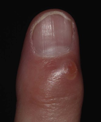
157 Myxoid cyst David de Berker Evidence Levels: A Double-blind study B Clinical trial ≥ 20 subjects C Clinical trial < 20 subjects D Series ≥ 5 subjects E Anecdotal case reports Myxoid cysts are also known as digital mucus cysts or pseudocysts and represent a ganglion of the distal interphalangeal joint. They arise in different forms in soft tissues, typically above or distal to the distal interphalangeal joint. The most common presentation is as a translucent nodule on the dorsum of the digit. Management strategy Myxoid cysts contain gelatinous material that has escaped from the distal interphalangeal joint. Treatment can involve removal of the material combined with measures to prevent further escape from the joint. Simple drainage through an incision can achieve the first, but normally there is recurrence, with further synovial fluid in the myxoid cyst within a few weeks. Measures to prevent the further accumulation of fluid are directed at reducing joint pathology or blocking the pathway of escape of fluid. The first category includes the use of injected triamcinolone, which might reduce synovial inflammation and the pressure of fluid within the joint, and surgery for osteophytes. Osteophytes appear to weaken the joint capsule and possibly contribute to joint pathology and synovial fluid production. Their removal is associated with resolution of the myxoid cyst. Blockage of the path of fluid escape is achieved by a range of traumatic and scarring procedures. The challenge is to produce an effective scar over the joint without excess morbidity or a long-term nail dystrophy. A practical compromise of morbidity, complexity of treatment, and efficacy is to employ cryosurgery in the first instance. This is best used with a distal block using plain lidocaine or bupivacaine. The cyst is then incised and drained. Two 20-second freezes with liquid nitrogen are given, with a complete thaw in between. Over the next 2 weeks the wound is dressed. This results in success in about 50% of cases involving fingers. Those that fail can be re-treated in the same way or proceed to surgery. Morbidity is least when there is precise surgery to the path of fluid escape. This can be identified by methylene blue injection into the joint and then raising a flap in a distal to proximal path, containing the cyst in the roof of the flap. The pathway is dyed and can be tied with absorbable ligature. The flap is sutured back in place and success is reported as 94% in the fingers. In some instances patient preference or medical considerations may mean that surgery is the first-line therapy. Specific investigations Transillumination Pricking and expression of gelatinous material Ultrasound MRI Transillumination is a useful clinical aid. Where there is diagnostic doubt, simple incision can be helpful to demonstrate the gelatinous material. Alternatively, high-resolution ultrasound or MRI can be employed. The first can only help define whether the structure is cystic or not. The latter provides better anatomical definition, and in over 80% of cases allows location of the pedicle communicating between the cyst and joint. Plain X-ray may reveal osteophytes of osteoarthritis, but this will not usually alter treatment. Benign tumors and pseudotumors of the nail: a novel application of sonography. Wortsman X, Wortsman J, Soto R, Saavedra T, Honeyman J, Sazunic I, et al. J Ultrasound Med 2010; 29: 803–16. MR imaging of digital mucoid cysts. Drapé JL, Idy Peretti I, Goettmann S. Radiology 1996; 200: 531–6. Only gold members can continue reading. Log In or Register to continue Related Related posts: Cat scratch disease Hemangiomas Tinea capitis Herpes genitalis Necrolytic migratory erythema Nevoid basal cell carcinoma syndrome Stay updated, free articles. Join our Telegram channel Join Tags: Treatment of Skin Disease Comprehensive Therapeutic Strategies Aug 7, 2016 | Posted by admin in Dermatology | Comments Off on Myxoid cyst Full access? Get Clinical Tree
157 Myxoid cyst David de Berker Evidence Levels: A Double-blind study B Clinical trial ≥ 20 subjects C Clinical trial < 20 subjects D Series ≥ 5 subjects E Anecdotal case reports Myxoid cysts are also known as digital mucus cysts or pseudocysts and represent a ganglion of the distal interphalangeal joint. They arise in different forms in soft tissues, typically above or distal to the distal interphalangeal joint. The most common presentation is as a translucent nodule on the dorsum of the digit. Management strategy Myxoid cysts contain gelatinous material that has escaped from the distal interphalangeal joint. Treatment can involve removal of the material combined with measures to prevent further escape from the joint. Simple drainage through an incision can achieve the first, but normally there is recurrence, with further synovial fluid in the myxoid cyst within a few weeks. Measures to prevent the further accumulation of fluid are directed at reducing joint pathology or blocking the pathway of escape of fluid. The first category includes the use of injected triamcinolone, which might reduce synovial inflammation and the pressure of fluid within the joint, and surgery for osteophytes. Osteophytes appear to weaken the joint capsule and possibly contribute to joint pathology and synovial fluid production. Their removal is associated with resolution of the myxoid cyst. Blockage of the path of fluid escape is achieved by a range of traumatic and scarring procedures. The challenge is to produce an effective scar over the joint without excess morbidity or a long-term nail dystrophy. A practical compromise of morbidity, complexity of treatment, and efficacy is to employ cryosurgery in the first instance. This is best used with a distal block using plain lidocaine or bupivacaine. The cyst is then incised and drained. Two 20-second freezes with liquid nitrogen are given, with a complete thaw in between. Over the next 2 weeks the wound is dressed. This results in success in about 50% of cases involving fingers. Those that fail can be re-treated in the same way or proceed to surgery. Morbidity is least when there is precise surgery to the path of fluid escape. This can be identified by methylene blue injection into the joint and then raising a flap in a distal to proximal path, containing the cyst in the roof of the flap. The pathway is dyed and can be tied with absorbable ligature. The flap is sutured back in place and success is reported as 94% in the fingers. In some instances patient preference or medical considerations may mean that surgery is the first-line therapy. Specific investigations Transillumination Pricking and expression of gelatinous material Ultrasound MRI Transillumination is a useful clinical aid. Where there is diagnostic doubt, simple incision can be helpful to demonstrate the gelatinous material. Alternatively, high-resolution ultrasound or MRI can be employed. The first can only help define whether the structure is cystic or not. The latter provides better anatomical definition, and in over 80% of cases allows location of the pedicle communicating between the cyst and joint. Plain X-ray may reveal osteophytes of osteoarthritis, but this will not usually alter treatment. Benign tumors and pseudotumors of the nail: a novel application of sonography. Wortsman X, Wortsman J, Soto R, Saavedra T, Honeyman J, Sazunic I, et al. J Ultrasound Med 2010; 29: 803–16. MR imaging of digital mucoid cysts. Drapé JL, Idy Peretti I, Goettmann S. Radiology 1996; 200: 531–6. Only gold members can continue reading. Log In or Register to continue Related Related posts: Cat scratch disease Hemangiomas Tinea capitis Herpes genitalis Necrolytic migratory erythema Nevoid basal cell carcinoma syndrome Stay updated, free articles. Join our Telegram channel Join Tags: Treatment of Skin Disease Comprehensive Therapeutic Strategies Aug 7, 2016 | Posted by admin in Dermatology | Comments Off on Myxoid cyst Full access? Get Clinical Tree