Mycobacterial (atypical) skin infections


Mycobacterial (atypical) skin infections


Ure Eke and John Berth-Jones


Evidence Levels:  A Double-blind study  B Clinical trial ≥ 20 subjects  C Clinical trial < 20 subjects  D Series ≥ 5 subjects  E Anecdotal case reports


image


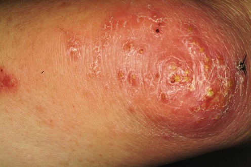
Fish tank (swimming pool) granuloma


Fish tank granuloma is an infection of the skin caused by Mycobacterium marinum, causing plaques and nodules, commonly on the upper extremities, which may spread in a sporotrichoid fashion after an incubation period of 2 to 6 weeks. The most common sources of infection are tropical fish aquariums, and swimming pools. The infection is commonly limited to the skin, but tenosynovitis, osteomyelitis, and arthritis have been reported with deeper infections. Rarely, disseminated infection may occur, especially in the immunocompromised. There are reports of the development of cutaneous M. marinum infection whilst on infliximab and adalimumab although paradoxically, infliximab has been found to be a useful adjunctive therapy in the management of M. marinum infection.


Management strategy


No controlled trials have been conducted, probably owing to the paucity of cases. Successful treatment is reported with antibiotics given singly or in combination. The mean duration of treatment in various reports ranges from 6 to 20 weeks. No statistical difference in efficacy between treatments has been demonstrated. Lesions can often be effectively treated by simple excision, but occasionally this seems to result in a prolonged infection. Heat treatment of the infected area may have an adjunctive role. Although spontaneous resolution may occur within three years, treatment is required for rapid recovery from infection and the prevention of dissemination. The second-line treatments described below have been so designated as there is relatively little published information about them. The third-line treatments are probably best regarded as adjunctive.


Specific investigations


Histology shows non-caseating granulomas, but a complete absence of epithelioid cells and multinucleate giant cells is not unusual in acute lesions. Suppurative, tuberculoid and palisading patterns of granulomas have been described. Dermal small vessel proliferation with mixed inflammation may be a good indicator of cutaneous atypical mycobacterial infections. Ziehl–Nielsen staining is positive for acid-fast bacilli in 30% of biopsies. Culture of the biopsy specimen at 30–33°C yields pigmented colonies of M. marinum. PCR can provide rapid and sensitive detection of mycobacterial DNA in formalin-fixed, paraffin-embedded specimens. In vitro sensitivity studies have not been uniformly predictive of clinical response to the antibiotics. Although they do not have a routine role in directing initial treatment, they may be useful in resistant cases.


First-line therapies


Epidemiological, clinical, and therapeutic pattern of Mycobacterium marinum infection: a retrospective series of 35 cases from southern France.


Eberst E, Dereure O, Guillot B, Trento C, Terru D, van de Perre P, Godreuil S. J Am Acad Dermatol 2012; 66: e15–16.


In this retrospective series, the treatment of 35 consecutive cases of M. marinum was reviewed. Thirty-four of 35 (97%) had complete clearance of skin lesions with one course of oral antimicrobial therapy given over a period of four to 24 weeks (median 12.4 weeks). Single antibiotic therapy was given successfully in 25 patients. Fourteen of these received minocycline 200 mg od, five patients received doxycycline 200 mg od and six patients received clarithromycin 500 mg od. Nine patients had dual antibiotic therapy using a combination of clarithromycin with doxycycline, minocycline, rifampicin or ofloxacin.


Third-line therapies


Stay updated, free articles. Join our Telegram channel

Aug 7, 2016 | Posted by in Dermatology | Comments Off on Mycobacterial (atypical) skin infections

Full access? Get Clinical Tree

Get Clinical Tree app for offline access