Fig. 2.1
Nerves of abdominal wall
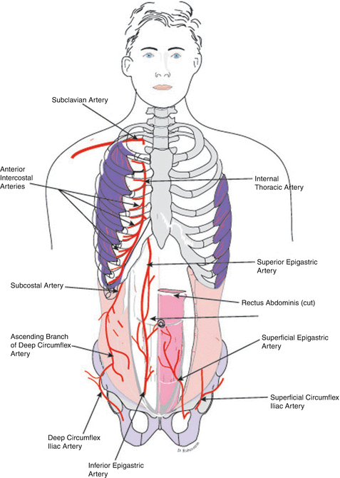
Fig. 2.2
Arteries of the abdominal wall
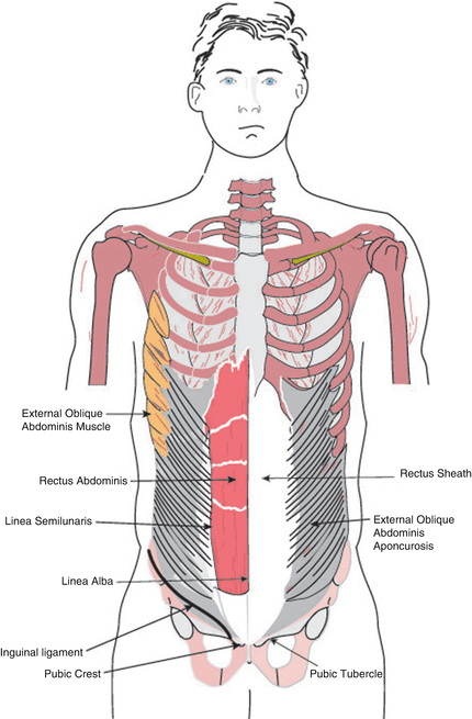
Fig. 2.3
The external abdominal oblique muscle, aponeurosis, and inguinal ligament
It has been concluded from a study of the arterial blood supply of the external oblique that the cranial part of this muscle is supplied by the posterior intercostal arteries, whereas the caudal part of the muscle received its primary blood supply from the deep circumflex iliac artery and to a limited degree from the iliolumbar artery. It has also been shown that the lateral branches of the deep circumflex iliac and iliolumbar arteries course on the external surface, while the anterior branches pierce the muscle from its posterior surface [19]. The principal role of the deep circumflex iliac artery in the blood supply of the muscle has been confirmed by an arterial injection research study conducted by Kuzbari et al. [20].
2.6 External Abdominal Oblique Muscle and Aponeurosis and Inguinal Ligament (Fig. 2.3)
The muscular part of the external abdominal oblique continues as aponeurosis at the lateral border of the rectus abdominis. The aponeurosis covers the anterior surface of the rectus abdominis, contributing to the anterior layer of the rectus sheath. It joins the aponeuroses of the internal oblique and the transverse abdominis to form the linea alba. The latter is a midline tendinous raphe that extends between the xiphoid process and the symphysis pubis and pubic crest. The adminiculum linea alba refers to the triangular part of the linea alba that attaches to the pubic crest.
The supraumbilical part of the linea alba is markedly wider and separates the recti completely, whereas the infraumbilical part is narrower and has a less distinct demarcation. Avascularity of the linea alba allows it to be used as a good site for surgical incisions. The lateral border of the rectus abdominis extending from the costal arch at or near the 9th costal cartilage to the pubic tubercle marks the semilunar (Spigelian) line. The latter also marks the sites of entry of the intercostal nerves into the rectus abdominis, rendering surgical incisions at this site unadvisable. The junction of the linea semilunaris and arcuate line of Douglas (transition point between the upper two thirds and the lower one third of the posterior layer of the rectus sheath) can be the site of the Spigelian hernia, which consists of extraperitoneal fat covered by the skin, superficial fascia, and aponeurosis of the external oblique.
Backward and slightly upward enfolding of the aponeurosis of this muscle between the anterior superior iliac spine and the pubic tubercle forms the inguinal (Poupart) ligament, which separates the site of inguinal from that of the femoral hernia. It also marks the transition between the abdominal wall and thigh. Its curved border constitutes the floor of the inguinal canal and Hesselbach’s triangle and maintains an oblique angle to the horizontal.
2.7 Aponeurosis of External Oblique, Superficial Inguinal Ring, and Inguinal Ligament (Fig. 2.4)
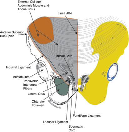
Fig. 2.4
The aponeurosis of the external oblique, the superficial inguinal ring, and the inguinal ligament
Examination of the posterior surface of the lateral crus of the superficial inguinal ring reveals a reflected part of the inguinal ligament, which is formed by the fibers of the external oblique aponeurosis that course superomedially to join the rectus sheath and linea alba anterior to the conjoint tendon. A posterolateral triangular extension from the medial part of the inguinal ligament that attaches to the medial end of the pecten pubis is known as the lacunar (Gimbernat) ligament or the pectineal part of the inguinal. This triangular ligament is separated from the femoral vein by the femoral canal, a potential site of femoral hernia. An extension of the fascia lata of the thigh that joins the inguinal ligament, the pectineal fascia, the periosteum of the pecten pubis, and the fibers of the transversalis fascia forms the fascial lacunar ligament. It is approximately 1 cm anterior and inferior to the pecten pubis and 3 cm lateral to the pubic tubercle. This ligament contributes to the femoral sheath which invests the femoral vein and artery.
The firm fibrous band that extends laterally along the sharp edge of the pecten pubis connecting the base of the lacunar ligament to the pecten pubis is known as the Cooper’s ligament. This ligament is further strengthened by the fibers that are derived from the pectineal fascia and adminiculum alba. The significant role of this thickening of the pectineal fascia in laparoscopic surgery of the inguinal region and female urinary incontinence has been reported by Faure et al. [21] and Rousseau et al. [22]. The Cooper’s ligament is anchored to the transversalis fascia in McVay’s technique of repair of inguinal hernia [23].
The gap that lies posterior to the inguinal ligament that allows the passage of vessels and nerves that supply the thigh is divided into vascular (lacuna vasorum) and muscular (lacuna musculorum) compartments. The dividing septum, the iliopectineal arch, is continuous with the iliopsoas fascia and inguinal ligament. The femoral vein and artery surrounded by the femoral sheath and the medially positioned femoral ring are located within the vascular compartment, whereas the muscular compartment contains the femoral nerve and iliopsoas muscle. Superior to the inguinal ligament and superolateral to the pubic tubercle, the superficial inguinal ring makes its appearance as a gap within the external oblique aponeurosis.
Despite some variations, the superficial inguinal ring is smaller in the female due to the relative thinness of the round ligament. However, it usually remains confined to the medial third of the inguinal ligament. The superficial inguinal ring has a stronger outer border formed by the lateral crus, which attaches to the pubic tubercle; an inner thin border formed by the medial crus, which interdigitates at the symphysis pubis; and a base at the pubic crest. The apex is formed by the intercrural fibers that interconnect the crura and thus resist the undue widening of the superficial inguinal ring. The spermatic cord in the male and the round ligament in the female exist at the superficial inguinal ring across the lateral crus invested by the external spermatic fascia, a continuation of the external abdominal aponeurosis. The external oblique muscle is innervated by the ventral primary rami of the lower five or six intercostal nerves.
Deep to the external oblique muscle, a much thinner internal abdominal oblique (Fig. 2.5) muscle lies, originating from the lateral two thirds of the inguinal ligament and iliac crest and sharing origin with the latissimus dorsi from the thoracolumbar fascia. Fibers of this muscle, particularly those from the iliac crest and thoracolumbar fascia, follow an upward and lateral course perpendicular to that of the external abdominal oblique muscle fibers, attaching to the lower borders of the lower three or four ribs and continuing with the internal intercostal muscles. The internal oblique becomes aponeurotic at the lateral border of the rectus abdominis and forms the linea alba by joining the aponeuroses of the ipsilateral and contralateral flat abdominal muscles.
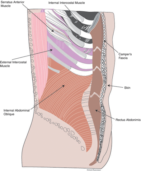

Fig. 2.5
Deep to the external oblique muscle, a much thinner internal abdominal oblique
The aponeurosis of the part of this muscle derived from the inguinal ligament arches over the spermatic cord in the male or the round ligament in the female to form the conjoint tendon (falx inguinalis) by uniting with the aponeurosis of the transverse abdominis muscle anterior to the rectus abdominis muscle. This tendon attaches to the pubic crest and to the medial part of the pecten pubis and is sutured to the transversalis fascia and to the reflected part of the inguinal ligament in the Bassini technique of herniorrhaphy [24–27]. The part of the conjoint tendon that attaches to the pecten pubis lies posterior to the superficial inguinal ring, forming a natural barrier that may resist the development of inguinal hernia. Due to the location of this tendon posterior to the superficial inguinal ring, a direct inguinal hernial pouch may protrude through acquiring the aponeurotic structure of this tendon.
Despite the variations in the attachment and structural characteristics, the conjoint tendon frequently joins the anterior layer of the rectus sheath medially and the interfoveolar ligament, a variable fibrous cord that connects the transverse abdominis to the superior pubic ramus, laterally. In the upper two thirds of the rectus sheath, defined by the part superior to the arcuate line or superior to the midpoint between the umbilicus and the symphysis pubis, the internal oblique aponeurosis splits into two layers. The anterior layer covers the anterior surface of the rectus abdominis joining the aponeurosis of the external oblique whereas the posterior layer invests the posterior surface of the rectus abdominis and joins the aponeurosis of the transverse abdominis and the transversalis fascia. In the lower third of the rectus sheath distal to the arcuate line or distal to the midpoint between the umbilicus and the symphysis pubis, the aponeurosis of the internal oblique remains a single layer and contributes to the anterior layer of the rectus sheath by joining the aponeuroses of the external oblique and transverse abdominis.
The internal oblique forms the lateral border of the Grynfeltt’s triangle, a potential site for superior lumbar hernia. This triangular space is bounded superiorly by the 12th rib and the serratus posterior inferior and by the erector spinae medially [28].
The loosely structured and sparsely arranged fibers of the internal oblique muscle and its aponeurosis extend around the spermatic cord and testis to form the cremasteric muscle and fascia. Fibers of the transverse abdominis usually contribute to the cremasteric, a striated muscle with an external and an internal part separated by the internal spermatic fascia [29]. The thin inconstant internal part originates from the pubic tubercle, conjoint tendon, and possibly the transverse abdominis, whereas the thick external part is directly derived from the inguinal ligament and its attachment to the anterior superior iliac spine. This involuntary muscle receives motor innervation from the genital branch of the genitofemoral nerve, which originates from the L1 to L2 spinal segments. The cremasteric muscle and fascia coil over the spermatic cord and testis at the inferior border of the internal oblique to join the anterior layer of the rectus sheath at the pubic tubercle. It has been reported that cautious dissection of the cremasteric muscle and fascia greatly enhances the exposure of the inguinal canal and deep inguinal ring in hernia repair [30]. In the female, the sporadic fibers of the external part of the cremasteric muscle wrap around the round ligament. Contraction of the cremasteric muscle occurs in the cremasteric reflex, a brisk reflex elicited by stimulation of the upper medial thigh, producing elevation of the testicles toward the superficial inguinal ring. Research studies have focused on the association between testicular torsion and hyperactive cremasteric reflex, and worsening of this condition in cold weather has been documented [31]. The internal abdominal oblique receives innervation from the ventral primary rami of the lower six intercostal nerve, as well as the iliohypogastric and ilioinguinal nerves.
Contraction of the external abdominal oblique muscles synchronously with the internal oblique and rectus abdominis muscles produces flexion of the vertebral column. Ipsilateral contraction of the external and internal abdominal oblique muscles causes ipsilateral abduction or lateral flexion of the trunk. Contraction of the ipsilateral external abdominal oblique together with the contralateral internal oblique results in rotation of the lumbar vertebral column.
The most posterior muscular layer of the anterior abdomen is the transverse abdominis, a relatively thin and wide muscle that consists of fibers that course nearly horizontal deep to the internal abdominal oblique muscle (Fig. 2.6). Like the internal oblique, it derives its fibers from the iliac crest, inguinal ligament, and thoracolumbar fascia but with some additional origin from the inner surfaces of the lower five or six ribs, partly interdigitating with the origin of the peripheral muscular part of the diaphragm. It may be absent or combined with the internal abdominal oblique and may be disrupted by several gaps filled with fascia. This muscle becomes aponeurotic near the lateral border of the rectus abdominis and joins the aponeuroses of other abdominal muscles to form the rectus sheath and the linea alba. An exception to this is at the level of the xiphoid process where muscle of the transverse abdominis follows a course deeper to the rectus abdominis and becomes aponeurotic much more medially near the linea alba.
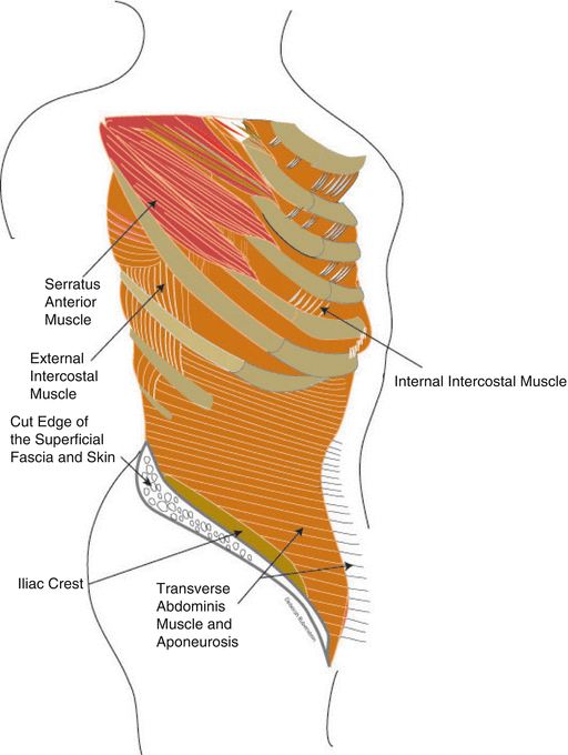

Fig. 2.6
Transverse abdominis muscle and aponeurosis
In the upper two thirds of the rectus sheath, which corresponds to the part superior to the arcuate line (midpoint between the umbilicus and symphysis pubis), the aponeurosis of the transverse abdominis contributes to the posterior layer of the rectus sheath by joining the posterior layer of the internal oblique aponeurosis and the transversalis fascia. In the lower one third of the rectus sheath, which corresponds to the part of the rectus sheath inferior to the arcuate line (midpoint between the umbilicus and symphysis pubis), the transverse abdominis aponeurosis contributes to the anterior layer of the rectus sheath by joining the aponeuroses of the oblique abdominal muscle. The lower fibers of the aponeurosis of this muscle contribute to the formation of the conjoint tendon by curving downward and medially above the spermatic cord in the male and the round ligament in the female and joining the aponeurosis of the internal abdominal oblique at the pubic crest.
The Spigelian hernia refers to a well-defined defect in the aponeurosis of the transverse abdominis through the semilunar (Spigelian) line at the lateral border of the rectus abdominis (Spigelian aponeurosis). The hernial sac, surrounded by extraperitoneal fat, is often interparietal and not easily palpable or diagnosed due to its location posterior to the aponeurosis of the external oblique [32]. Irrespective of the age at the time of manifestations, this hernia can be congenital of origin, commonly seen above the arcuate line in the lower abdomen, and develops synchronously with inguinal hernias in neonates [33]. Acquired Spigelian hernia can be due to obesity, previous surgery, scarring, or as a complication of ambulatory care dialysis, ambulatory peritoneal dialysis, and hernial complications [34]. Patients with this rare type of hernia may present with localized pain or signs of intestinal obstruction due to strangulation.
This muscle is innervated by the ventral rami of the lower five or six intercostal nerves, as well as by the subcostal, iliohypogastric, and ilioinguinal nerves. In general, the innervation of the flat abdominal muscle, particularly by the intercostal nerves, shows extensive networking and considerable overlap. This fact may explain the limited or complete absence of detectable deficits upon damage to one or two intercostal nerves. In contrast, the rectus abdominis is segmentally innervated and lacks or shows very limited overlap, and as a result, individual intercostal nerve damage is most likely to cause segmental rectus abdominis deficit instead of total muscle dysfunction.
Stay updated, free articles. Join our Telegram channel

Full access? Get Clinical Tree


