Early complications
Late complications
Seroma
Dog ears
Cellulitis
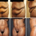
Lateral fat excess
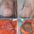
Wound dehiscence and necrosis
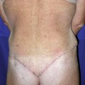
Scar and contour asymmetry
Abscess and surgical site infections
Seroma
Hematoma
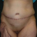
Umbilical stenosis or dissatisfaction
Pulmonary emboli
Neuropathic pain
39.2.1 Seroma
Seroma is the most common complication after abdominoplasty, occurring in around 5–30 % of patients [5]. Differently from hematomas, which appear more suddenly and generally require a more aggressive treatment with surgical re-intervention in about 50 % of cases [2], seromas develop after the first postoperative week and are the most common source of infection after lower truncal procedures [6]. Even if they may spontaneously resolve, multiple aspirations are usually required, with increased frequency of follow-up and possible development of infection, which cause significant disability by delaying recovery times and impairing normal wound healing [7]. In a retrospective analysis of complications of over 200 abdominoplasty procedures [2], seromas had to be aspirated in 40 % of the cases for an average of 2.5 times, which is consisted to other published works [8], with final retake to the OR in about 5 % of the cases. All this makes seroma a particularly feared complication in the practice of the aesthetic surgeon.
39.2.1.1 Pathophysiology of Seroma
Seroma is a fluid collection, rich in neutrophils and protein, and with the characteristics of an exudate [6, 9]. The etiology is not completely understood and is probably multifactorial. Among the mechanisms involved in seroma formation, dissection, detachment, and shearing of fasciocutaneous flaps [6, 10] with consequent damage of lymphatic architecture seem to be the key etiologic factors [4]. The loss of anchorage of the abdominal skin flap and the rectus sheath results in a dead space where fluid can drain and collect. If small fluid collections can be spontaneously reabsorbed, bigger amounts may increase pressure causing wound dehiscence, necrosis, and infection, which result in significant morbidity and delay in recovery [4, 11].
39.2.1.2 Patient Features and Risk Factors
Main patient’s predisposing factors associated to seroma development are obesity (BMI > 30 kg/m2) and massive weight loss [5, 6]. Coon et al. [12] reported significantly increased complications in the post-bariatric population, similarly to Neaman [2] in a retrospective review of abdominoplasty complications. Incidence of seroma in these patients is significantly higher, reaching almost 40 % [13]. Diabetes, smoking, and hypertension have been found to increase general complication rates after abdominoplasty, acting through the microvascular damage [2, 14]. Even if these factors may predispose to seroma formation, there is no evidence-based proof of direct effect on seroma formation. In our current practice, obese and post-bariatric patients are routinely informed prior to operation of the relevant risk of complications. Smokers are asked to discontinue or decrease smoking 1 month in advance.
39.2.1.3 Surgical Approaches to Prevent Seroma
Different surgical approaches have been used to prevent postoperative seroma formation, some of them showing scientific evidence. The level of dissection of the abdominal flap has been examined as having an effect on seroma formation and drain maintenance [1]. Traditionally, the abdominal flap raising was carried out on the anterior muscle fascia. The first technical trend is to limit flap dissection over the umbilicus with limited undermining up to the xiphoid process, resulting in reduced dead space [6]. The second key modification is to preserve Scarpa’s fascia and deep adipose tissue in the infraumbilical region. These refinements allow preserving in both the superficial and deep lymphatic system (located in the dermis and in the deep adipose tissue adjacent to the Scarpa’s fascia, respectively) [6, 15]. Particularly, Scarpa’s fascia preservation has shown to highly significantly reduce drainage output and seroma formation down to 0–2.5 % [16, 17].
Another technical refinement to prevent seromas is progressive tension or quilting sutures to fix the abdominal flap to the rectus fascia [18, 19]. These technical modifications all refer to the basic concept of minimizing abdominal flap friction and increase adherence to the deep fascia. At the same time, tension is dispersed along the flap and not on the suture line [1]. Marsh et al. [5] showed that additional flank liposuction procedures could significantly increase in seroma formation (36 % vs 8 % in controls without liposuction), potentially by creating supplementary dead space through the tunneling created by the aspiration cannulas and consistently to what found in other reports [8, 20]. However, the literature on the effect of liposuction is inconsistent as many other studies did not find differences in seroma formation [6, 13]. Besides statistical significance, what comes out is that liposuction of the flanks is often a necessary procedure to avoid lateral fat excess and generally patients experience the highest satisfaction from these combined procedures [21]. The theoretical increased seroma risk may probably be reduced following the basic technical principles abovementioned, such as limited central abdominal undermining and maintenance of deep abdominal fat layer [1]. Even if it has been argued that it is not necessary to place drains after quilting sutures [22], the authors use two drains in full abdominoplasty procedures, with removal when output is <40 mL/24 h. Active lymphatic drainage is started on postoperative day 1 in patients at particular high risk (obese patients and massive weight loss). In these patients, the use of fibrin glues may be a useful supplementary tool. Fibrin seals microvascular and connective tissue injuries on wound surfaces, reducing the dead space where seromas tend to form and significantly decreasing drainage output and hospital stay [7, 10]. Drains are avoided in miniabdominoplasties, low-BMI patients, or when fibrin glue has eventually been used. However, drains seem to be a rather personal decision of the surgeon, and no systematic literature is present on the argument.
39.2.1.4 Treatment of Early and Late Complications Affecting the Aesthetic Outcomes
Once established, seromas can expand and consequently increase the pressure under the abdominal flap, leading to dehiscence, spontaneous drainage, or infection. Seroma pockets that become infected often present with overlying cellulitis, fever, and rising inflammatory parameters. Collected fluids may or may not spontaneously drain. In both cases, a microbiologic sample is taken of the discharge (by direct aspiration or under ultrasound guidance), leaving a drain in place. Intravenous antibiotic therapy by amoxicillin-clavulanic acid is empirically started and secondarily adapted according to antibiogram results. If signs of sepsis are present or the collection is considered too big to be drained exclusively by percutaneous interventions, aggressive surgical drainage is generally required, followed by the antibiotic IV therapy abovementioned.
Concerning noninfected recurrent seromas, the usual initial treatment consists of repeated aspirations. This requires multiple visits to the surgeon’s office, increasing postoperative costs and patient’s discomfort [6
 Abdominoplasty Combined with Cesarean Section: Discussion of the Evidence
Retrospective Analysis of Never Events in Panniculectomy and Abdominoplasty Patients and Their Financial Implications
Abdominoplasty Combined with Cesarean Section: Discussion of the Evidence
Retrospective Analysis of Never Events in Panniculectomy and Abdominoplasty Patients and Their Financial Implications
 No Drain Abdominoplasty with Progressive Tension Suture: The Logic, Simplicity, and Aesthetics
 Abdominoplasty as the Impetus for Selection of the Deep Inferior Epigastric Artery Perforator Free Flap for Non-breast Reconstruction
 Circumferential Abdominoplasty
 Abdominal Wall Repair Post Hernia in Kidney and Liver Transplantation


Related posts:
Abdominoplasty Combined with Cesarean Section: Discussion of the Evidence
Retrospective Analysis of Never Events in Panniculectomy and Abdominoplasty Patients and Their Financial Implications
No Drain Abdominoplasty with Progressive Tension Suture: The Logic, Simplicity, and Aesthetics
Abdominoplasty as the Impetus for Selection of the Deep Inferior Epigastric Artery Perforator Free Flap for Non-breast Reconstruction
Circumferential Abdominoplasty
Abdominal Wall Repair Post Hernia in Kidney and Liver Transplantation
Stay updated, free articles. Join our Telegram channel
Full access? Get Clinical Tree
