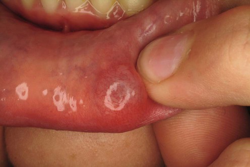
151 Mucoceles Noah Scheinfeld Evidence Levels: A Double-blind study B Clinical trial ≥ 20 subjects C Clinical trial < 20 subjects D Series ≥ 5 subjects E Anecdotal case reports Labial mucocels fall into two categories: the mucous extravasation cyst, and the mucous retention cyst. The mucous extravasation cyst describes a false cyst because the mucous extravasation cyst lacks an epithelial lining arising from the partially or totally severed salivary gland duct resulting in the accumulation of saliva in the adjacent soft tissue. At this point the mucocele is cut off by a fibrous connective tissue pseudocapsule. The ductal epithelium lines the mucous retention cyst. The mucous retention cyst develops from partial obstruction of a duct in the presence of the salivary gland’s continued mucous secretion. The extravasation mucocele manifests most commonly and manifest most often on the young person’s lower lip. The retention mucocele is more apt to occur on the buccal cheek or soft palate of an older patient. To expand this further, mucous extravasation cysts arise from trauma to salivary gland ducts. This trauma leads to rupture salivary gland ducts and leakage of mucin from the minor salivary glands. The mucin subsequently forms pseudocytic aggregations most commonly on the lower lip. These aggregations are referred to as mucoceles. Mucoceles manifest with a variety of tones and color that range from flesh color to red to translucent blue. The shape of mucoceles is round or oval and their surface is smooth. They usually possess a soft, fluctuant or gel-like consistency. Single or multiple mucoceles can manifest and can range from 0.1 to 2 cm mm in diameter. The natural history of mucoceles can involvement their expansion and periodic rupture and sometimes spontaneous resolution. There is some morbidity associated with mucocles that ranges from discomfort, to suboptimal cosmetic to appearance of a nodule with a hardened consistency due to scarring and tissue consolidation. ‘Superficial mucocele’, a variant of a mucocele, can manifest on the palate, retromolar pad, and posterior buccal mucosa. Superficial mucocles manifest as single or multiple vesicles, which can break down into an ulcer. Despite healing after a few days, superficial mucoceles recur often in the same location. A mucocele is termed a ranula when on the floor of the mouth, and epulis when on the gums. Specific investigations Biopsy Doppler ultrasonography Color Doppler imaging First-line therapies Cryotherapy C Intralesional corticosteroids D No treatment (observation) D Only gold members can continue reading. Log In or Register to continue Related Related posts: Cat scratch disease Hemangiomas Tinea capitis Herpes genitalis Necrolytic migratory erythema Nevoid basal cell carcinoma syndrome Stay updated, free articles. Join our Telegram channel Join Tags: Treatment of Skin Disease Comprehensive Therapeutic Strategies Aug 7, 2016 | Posted by admin in Dermatology | Comments Off on Mucoceles Full access? Get Clinical Tree
151 Mucoceles Noah Scheinfeld Evidence Levels: A Double-blind study B Clinical trial ≥ 20 subjects C Clinical trial < 20 subjects D Series ≥ 5 subjects E Anecdotal case reports Labial mucocels fall into two categories: the mucous extravasation cyst, and the mucous retention cyst. The mucous extravasation cyst describes a false cyst because the mucous extravasation cyst lacks an epithelial lining arising from the partially or totally severed salivary gland duct resulting in the accumulation of saliva in the adjacent soft tissue. At this point the mucocele is cut off by a fibrous connective tissue pseudocapsule. The ductal epithelium lines the mucous retention cyst. The mucous retention cyst develops from partial obstruction of a duct in the presence of the salivary gland’s continued mucous secretion. The extravasation mucocele manifests most commonly and manifest most often on the young person’s lower lip. The retention mucocele is more apt to occur on the buccal cheek or soft palate of an older patient. To expand this further, mucous extravasation cysts arise from trauma to salivary gland ducts. This trauma leads to rupture salivary gland ducts and leakage of mucin from the minor salivary glands. The mucin subsequently forms pseudocytic aggregations most commonly on the lower lip. These aggregations are referred to as mucoceles. Mucoceles manifest with a variety of tones and color that range from flesh color to red to translucent blue. The shape of mucoceles is round or oval and their surface is smooth. They usually possess a soft, fluctuant or gel-like consistency. Single or multiple mucoceles can manifest and can range from 0.1 to 2 cm mm in diameter. The natural history of mucoceles can involvement their expansion and periodic rupture and sometimes spontaneous resolution. There is some morbidity associated with mucocles that ranges from discomfort, to suboptimal cosmetic to appearance of a nodule with a hardened consistency due to scarring and tissue consolidation. ‘Superficial mucocele’, a variant of a mucocele, can manifest on the palate, retromolar pad, and posterior buccal mucosa. Superficial mucocles manifest as single or multiple vesicles, which can break down into an ulcer. Despite healing after a few days, superficial mucoceles recur often in the same location. A mucocele is termed a ranula when on the floor of the mouth, and epulis when on the gums. Specific investigations Biopsy Doppler ultrasonography Color Doppler imaging First-line therapies Cryotherapy C Intralesional corticosteroids D No treatment (observation) D Only gold members can continue reading. Log In or Register to continue Related Related posts: Cat scratch disease Hemangiomas Tinea capitis Herpes genitalis Necrolytic migratory erythema Nevoid basal cell carcinoma syndrome Stay updated, free articles. Join our Telegram channel Join Tags: Treatment of Skin Disease Comprehensive Therapeutic Strategies Aug 7, 2016 | Posted by admin in Dermatology | Comments Off on Mucoceles Full access? Get Clinical Tree


 Cryotherapy
 Intralesional corticosteroids
 No treatment (observation)


