The initial evaluation of patients with morphea should involve: Active morphea lesions should be treated as the lesions are frequently symptomatic and can produce permanent cosmetic and functional sequelae. Initial evaluation should be focused on determining the extent, severity, and activity of morphea lesions laying the groundwork for rational therapeutic choices. Arkachaisri T, Vilaiyuk S, Torok KS, Medsger TA Jr. Rheumatology (Oxford) 2010; 49: 373–81.
Morphea
Management strategy
Initial evaluation
 Questions regarding recent development of new lesions/extension of existing lesions to determine level of activity
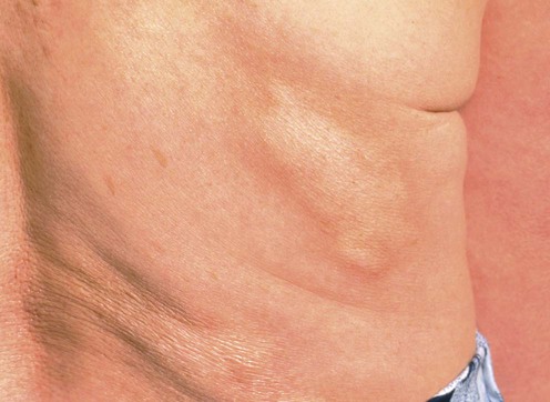
 Palpation of affected areas for depth of sclerosis
 Imaging with MRI if musculoskeletal involvement is suspected.
Specific investigations
Development and initial validation of the localized scleroderma skin damage index and physician global assessment of disease damage: a proof-of-concept study.
Related posts:
![]()
Stay updated, free articles. Join our Telegram channel

Full access? Get Clinical Tree