This article reviews the key anatomic structures in the region of the midface, including important surface and bony landmarks, innervation, blood supply, muscle layers, and fat compartments. It also discusses changes in these structures related to the aging process and aesthetic analysis of the midface to aid with operative planning.
Key points
- •
The midface is the region between the upper and lower thirds of the face. Within the midface there is an anterior portion referred to as the midcheek and the posterior portion referred to as the lateral cheek.
- •
The changes with aging tend to affect the midcheek structures with laxity of the soft tissue supporting ligaments (orbitomalar, zygomatic, and masseteric), bony atrophy, decreases in skin thickness elasticity, and subcutaneous fat resorption.
- •
Goals in midface rejuvenation are to produce midcheek fullness and smooth transition into adjacent areas of the lower lid and lower face.
Definition of the area
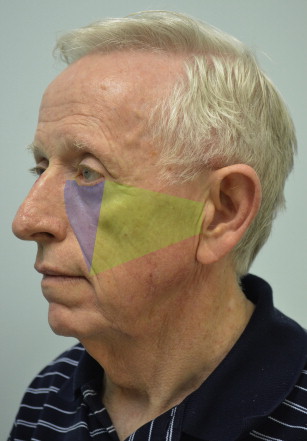
The midface is commonly used to describe the central third of the face because it is commonly divided into the upper, middle, and lower face. The upper border of the midface extends from the superior helix along the upper zygomatic arch to the lateral canthus and then along the lower lid to the nose. The lower border extends from the lower tragus to the oral commissure and along the nasolabial fold to the nose ( Fig. 1 ). The midface can further be divided by a line from the lateral canthus to the commissure. Anterior to the line is the midcheek and posterior is the lateral cheek.
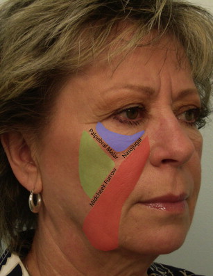
The midcheek and can further be divided into lid-cheek, malar, and nasolabial components ( Fig. 2 ). The palpebral malar crease separates the lower lid and malar fat divisions. The nasojugal crease separates the lower lid and nasolabial divisions. The midcheek furrow separates the malar and nasolabial divisions.
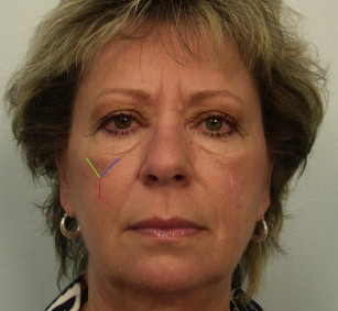
With aging, the midcheek divisions become apparent with development of a nasojugal fold medially, palpebral malar groove superolaterally, and a midcheek furrow inferolaterally in the shape of a Y in between the 3 components of the midcheek ( Fig. 3 ). The youthful midcheek typically blends into the lower lid, nose, nasolabial, and lateral facial regions without demarcation and has uniform fullness and volume.
With further anatomic study the superficial fat of the cheek itself has been shown to have 3 separate fat compartments: the medial, middle, and lateral temporal compartments, which all have separate septae. In addition, the sub–orbicularis oculi fat (SOOF) located in the lid-cheek division has also been shown to have 2 separate fat compartments. The medial component of the SOOF extends from the medial limbus to the lateral canthus along the orbital rim and the lateral component extends from the medial fat pad to the temporal fat pad.
Definition of the area
The midface is commonly used to describe the central third of the face because it is commonly divided into the upper, middle, and lower face. The upper border of the midface extends from the superior helix along the upper zygomatic arch to the lateral canthus and then along the lower lid to the nose. The lower border extends from the lower tragus to the oral commissure and along the nasolabial fold to the nose ( Fig. 1 ). The midface can further be divided by a line from the lateral canthus to the commissure. Anterior to the line is the midcheek and posterior is the lateral cheek.
The midcheek and can further be divided into lid-cheek, malar, and nasolabial components ( Fig. 2 ). The palpebral malar crease separates the lower lid and malar fat divisions. The nasojugal crease separates the lower lid and nasolabial divisions. The midcheek furrow separates the malar and nasolabial divisions.
With aging, the midcheek divisions become apparent with development of a nasojugal fold medially, palpebral malar groove superolaterally, and a midcheek furrow inferolaterally in the shape of a Y in between the 3 components of the midcheek ( Fig. 3 ). The youthful midcheek typically blends into the lower lid, nose, nasolabial, and lateral facial regions without demarcation and has uniform fullness and volume.
With further anatomic study the superficial fat of the cheek itself has been shown to have 3 separate fat compartments: the medial, middle, and lateral temporal compartments, which all have separate septae. In addition, the sub–orbicularis oculi fat (SOOF) located in the lid-cheek division has also been shown to have 2 separate fat compartments. The medial component of the SOOF extends from the medial limbus to the lateral canthus along the orbital rim and the lateral component extends from the medial fat pad to the temporal fat pad.
Internal organization/layers of the area
The layers in the midface are similar to those in the upper and lower face with skin, subcutaneous fat, a musculoaponeurotic layer, loose areolar layer, and periosteum/bone ( Figs. 4 and 5 ).
The bony framework consists of the zygoma and the maxillary bones with a small component of the lacrimal bone. Important attachments to the bony framework are the mimetic muscles, the zygomaticus major and minor, and the zygomatic ligament arising between and around the zygomaticus major and minor and minor muscles. There is limited bone available for attachment of soft tissue because the oral cavity mucosal reflexion occupies a large portion of the anterior maxilla. In addition, the prezygomatic space with the orbitomalar ligament superior and zygomatic ligaments inferiorly also limits direct soft tissue attachment to the zygomatic bone. This arrangement allows gliding of the soft tissues over the spaces and allows the separate functions of eye closure, smiling, and chewing.
In the midcheek region immediate superficial to the maxilla and zygoma lies the deep fat compartment, with preperiosteal fat and the buccal fat pad. This fat lies deep to the zygomaticus muscles and levator labii superioris, and within the fat pad are terminal branches of the zygomatic and buccal nerves that pass more superficially to innervate their muscle targets in the musculoaponeurotic layer. This layer allows gliding of the mimetic muscles (zygomaticus major and minor, levator labii superioris) with facial expressions. The zygomatic nerve innervates the zygomatic muscles on their deep surface but more medially innervates the levator superioris on its superficial surface.
The superficial musculoaponeurotic system (SMAS), often credited to Mitz and Peyronie, is a distinct layer in the face that is more developed in the lateral cheek region and becomes less distinct into the anterior face or midcheek region. Laterally the SMAS is more distinct separate from the parotid fascia and is continuous with the platysma inferiorly and the temporoparietal fascia superiorly. The SMAS also separates fat into a superficial fat compartment that contains septae and a deeper fat compartment without septae. In the midcheek region this is analogous to the SMAS/mimetic muscle layer separating the malar fat pad from the buccal fat pad. The exact relationship of the SMAS with the parotid fascia and mimetic muscles is not clearly shown in all studies, but most studies agree that the SMAS is most distinct in the parotid region and becomes thin and less substantial moving anterior into the midcheek region.
The subcutaneous layer in the midface includes the malar fat pad, which can be further subdivided into 3 separate compartments (medial, middle, and lateral), and each can age differently. Superior and deep to the malar fat pad is the SOOF, which occupies the prezygomatic space between the orbitomalar ligament and zygomatic ligament with the roof of the space being the orbicularis oculi muscle. The SOOF is separate and distinct from the malar fat and adherent to the deep surface of the orbicular oculi muscle, whereas the malar fat pad is superficial to the SMAS.
Related posts:

Stay updated, free articles. Join our Telegram channel

Full access? Get Clinical Tree


