Microtia represents a spectrum of maldevelopment of the external ear. Reconstructive techniques may utilize an autogenous rib cartilage framework and require 2–4 stages; alternatively, an alloplastic framework can be used and typically requires 1–2 stages. Successful reconstruction of microtia with either technique can provide a significant quality of life improvement, and both techniques are described in this article.
Key points
- •
Microtia, or abnormal external ear development, is a relatively rare congenital condition that is more common in certain ethnic groups, in men, and in the right ear.
- •
Given the complex structure of the ear and the difficulty in creating the anatomic environment for a neo-appendage, reconstruction of the auricle has always been a unique and challenging problem.
- •
There is no universal consensus on grading microtia. The most common nomenclature is the Weerda classification, which involves classifying the microtic ear on scale of grade 1 (small with normal features) to grade 3 (mass of deformed tissue).
- •
Autogenous rib reconstruction involves at least 2 stages (typically more), is generally a more durable reconstruction, and is less prone to infection.
- •
Alloplastic porous high-density polyethylene reconstruction typically involves 2 stages, is generally more aesthetic, involves less morbidity, and can be done at a younger age.
Videos of microtia reconstruction accompany this article at http://www.facialplastic.theclinics.com/
Overview
Microtia, or abnormal external ear development, occurs in 1 in 4000 to 10,000 births. It has a higher incidence in Asian, Hispanic, and Native American populations, with some studies citing a statistically significant increased risk in children of multiparous mothers. There is also a higher risk in males versus females, and microtia more commonly affects the right ear.
Embryologically, microtia is caused by malformation of the 6 hillocks that eventually join to form the auricle. During the sixth week of gestation, these hillocks form from the first and second branchial arches, eventually developing into the helix, lobule, tragus, and antihelix. The concha and external auditory meatus are formed by the first branchial groove and, as such, can be affected independently of the other structures.
Reconstruction of the auricle is a unique and challenging problem faced by surgeons today. The complex structure of the ear, along with the inherent difficulty of placing a framework within a tight skin pocket, leads to a spectrum of results among the varying surgeons who have performed these procedures. Over the years, methods of treatment have evolved, with techniques becoming more refined, but the core concepts remain the same. In general, there are 2 potential reconstructive options: autogenous rib cartilage and alloplastic implantation.
Historical perspective
Reports of ear reconstruction attempts date back to the sixteenth century, with the first documented successful reconstruction reported by Johann Friedrich Dieffenbach in the mid-nineteenth century, using a folded mastoid flap to repair a traumatic defect. Pierce discussed the use of a cartilage graft in 1930, with Gilles first describing attempted microtia reconstruction with donor cartilage from a patient’s mother in 1937. From this point forward, cartilage grafts (both human and bovine) gained favor; however, it was quickly noted that these grafts tended to soften and sag over time, with some ultimately resorbing or being rejected.
In 1943, Peer developed a technique whereby the auricle was prefabricated using costal cartilage fragments, which were fitted to a mold and stored in the abdomen for future implantation. There were issues surrounding the need for multiple operations, and that the structural integrity of the molded fragments could not withstand the deforming force of a tight skin pocket. Tanzer described the subcutaneous placement of an autogenous cartilage graft framework in 1959.
The history of alloplastic auricular reconstruction is more recent. The use of alloplastic implantation for auricular reconstruction was initially attempted in the 1960s using silicone implants, but this reconstruction technique was fraught with complications, with a high incidence of implant failure, especially related to minor trauma or abrasions. In 1990, Shanbhag and colleagues first wrote of the feasibility of using porous high-density polyethylene (PHDPE) in a baboon animal model. Wellisz and colleagues, in 1992, described the use of a prefabricated alloplastic implant for microtia reconstruction in humans, constructed from PHDPE, subsequently marketed in the United States under the trade name of Medpor PHDPE (Stryker, Kalamazoo, MI). This material proved to have many properties that are ideal for auricular reconstruction, and continues to be the alloplastic microtia reconstruction material of choice.
Patient assessment
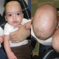
The grading of microtia suffers from the disagreement on a universally accepted standardized scale ( Table 1 ). The most commonly referenced scale was originally described by Weerda and later refined by Aguilar. The Weerda classification is based on the severity of auricular deformity: grade I describes a small ear with normal features, grade II describes a rudimentary auricle with some recognizable components, and grade III refers to a mass of deformed tissue. The Nagata grading is based according to vestigial structures rather than a scale.
| Classical | Atypical | |
|---|---|---|
| Brent | Remnant vestige (“sausage-shaped appendage”) Relatively normal lobule | All other types, including anotia, conchal remnants, vestiges with pits and grooves |
| Lobule Type | Conchal Type | Small Conchal Type | Anotia | |
|---|---|---|---|---|
| Nagata |
|
|
| Complete absence of an auricle |
| Grade I | Grade II | Grade III | |
|---|---|---|---|
| Weerda/Aguilar | Small ear: normal features | Rudimentary auricle: some recognizable components | Mass of deformed tissue |
Brent uses 2 general categories to describe microtia: classical and atypical. Classical is used to describe a vestige resembling a “sausage-shaped appendage” with a relatively normal lobule that is misplaced. The atypical category represents all other types, including anotia, microtias with conchal remnants, and vestiges with furrows, pits, or grooves.
Nagata also developed a grading system for microtia, consisting of the following categories: lobule type, concha type, small concha type, and anotia. Lobule type is a remnant of an ear with a lobule but no concha, acoustic meatus, or tragus. Concha type has some lobule, concha, acoustic meatus, tragus, and incisura tragica. Small concha type is a remnant ear and lobule with a small indent for a concha. Finally, anotia is the complete absence of an auricle.
Regardless of the system used to describe microtia, a surgeon must be mindful of other hypoplastic features of the face and temporal bone when planning reconstruction. Ultimately, symmetry is the goal.
Microtia is frequently associated with conductive hearing loss (CHL), with rates of concordant CHL upward of 90%. Sensorineural hearing loss is also seen, but at a much lower rate (∼15%). It is therefore critical to assess the extent of the external auditory canal development and the child’s hearing status. Children with bilateral CHL need to be fitted with a bone-conducting hearing aid from a young age to ensure normal cognitive development. Abnormalities associated with microtia include cleft lip/palate, microphthalmia, anophthalmia, cardiac defects, abnormal limb development, renal malformation, holoprosencephaly, and facial nerve dysfunction. Overt facial nerve dysfunction has been reported in as many as 15% of patients with microtia.
Associated anomalies occur in approximately 50% of cases and are most frequently in the regions arising from the first or second embryologic branchial arches. Craniofacial microsomia is a spectrum of malformations that affect these arches, and includes malformations such as Goldenhar syndrome, hemifacial microsomia, and oculoauricular vertebral dysplasia.
Microtia is more common in the setting of Goldenhar and Treacher Collins syndromes, among other, rarer syndromes. With this in mind, all newborns with a microtic ear should be fully assessed for concurrent congenital abnormalities.
Potential otologic surgery for atresia should be investigated. If aural atresia surgery is pursued, it is typically done after completed auricular reconstruction to preserve the blood supply for the reconstruction and aesthetic positioning of the neoauricle. A thorough physical examination should be performed with attention to facial development and animation, ear symmetry, and dental occlusion. Further otologic evaluation with an audiogram and high-resolution temporal bone computed tomography (CT) scan are indicated before surgery. The CT temporal bone provides important information regarding the status of the middle ear, ossicles, and course of the facial nerve, all critical when weighing the benefits of, or planning, otologic surgery. A lateralized facial nerve will be at risk of injury during otologic drill-out surgery for an atresia repair. Alternative measures, such as bone-anchored hearing aide (BAHA) placement, should be considered in this situation. Family history of genetic disorders should be elicited, and if appropriate, genetic testing should be initiated.
Timing of surgery plays an important role in reconstructive options. The age for optimal reconstruction varies depending on the particular surgical option.
In autogenous cartilage reconstruction, limitations in available rib cartilage must be balanced with potential psychological sequelae to a child with an auricular deformity. This has led to a general consensus that an appropriate window of rib reconstruction is between the ages of 7 and 10 years old. The Brent technique allows for the option for rib reconstruction as early as age 5, but the ideal age is still 7. The greater cartilage demands of the Nagata procedure require a larger chest circumference (at least 60 cm), which roughly correlates to an age of 10 for reconstruction. This limitation may be a factor for an anxious family or a child suffering psychological distress.
In comparison with autologous rib cartilage reconstruction, the indications for PHDPE reconstruction are narrower. PHDPE is indicated only in patients with Weerda second-degree and third-degree dysplasias, and in patients with failed autogenous rib reconstructions. PHDPE reconstruction can be done at an earlier age, as there is no need for donor rib to be a certain size. The patient should be at least 5 to 6 years old, as this is when the contralateral (normal) ear is approximately 85% of its normal, adult size. From a social perspective, this age also happens to coincide with the commencement of school, where the risk of ridicule and ostracism suddenly becomes higher.
Of equal importance is the number of stages of reconstruction a patient or family is willing to endure, as cartilage reconstructive options require a commitment to a greater number of surgeries than alloplastic reconstruction. It is also wise to consider the inherent maturity of the child, factoring in the child’s age and his or her observed behavior. Older children obviously have a greater understanding of what reconstruction entails and tend to be more engaged in the postoperative course. Familial concerns can be allayed with discussions and reassurance that the long-term benefits of a proper reconstruction ultimately outweigh the negatives of expedited surgery.
Current practice
Autogenous Cartilage
Rib reconstruction is the oldest method of microtia repair, and is touted by its proponents to be both the most stable in response to trauma, as well as more resistant to infection, when compared with alloplastic reconstruction. Essentially, rib reconstruction first involves carving and assembling a framework of costal cartilage with carefully placed sutures, and inserting this sculpted framework within a skin pocket in an aesthetically appropriate position. The later stages refine the appearance of the framework by introducing finer details, as well as elevating the framework away from the scalp.
The current era of the rib reconstruction has been credited to Tanzer. Other subsequent surgeons have contributed modifications and alterations to his technique, most notably Brent and Nagata. Regardless of specific technique, all reconstructive surgeries using rib cartilage adhere to the same general principles of tissue handling and skin redraping. The spectrum of techniques differs in terms of surgical timing, patient selection, and stage number.
Controversy continues to exist in terms of autogenous reconstruction itself, particularly between the Brent and Nagata techniques, the 2 most popular methods using costal cartilage. Both techniques borrow from Tanzer’s original description, but have their own advantages and disadvantages. Although the Nagata operation has fewer stages, and reconstruction has been demonstrated to be more consistent, this technique requires more cartilage, and thus can be performed only at an older age. The Brent operation emphasizes limited cartilage harvesting, along with minimization of the chest wall deformity. The reduced cartilage demand for this procedure means that children can begin surgery at a younger age, potentially limiting psychological sequelae at school.
To this effect, surgical planning is critical to all reconstructive efforts in the ear. Accurate assessment as to the structures present helps to guide incision design and framework crafting. Miscalculations will result in the suboptimal appearance of the reconstructed ear. The numerous modifications described in Nagata’s papers illustrate how attention to detail is necessary for creating a fabricated ear that is realistic and cosmetically appropriate; for example, although much of the Nagata procedure is the same for all 3, alterations in incisions and framework fabrication need to be customized to a patient’s preoperative microtia type.
Brent technique
The Brent technique is a 3-stage to 4-stage procedure using autogenous rib cartilage, and is recognized among various rib reconstructive techniques for its durability. As of 1999, Brent had reported more than 70 reconstructed ears that had survived major trauma. In addition to blunt trauma, there are reports of graft survival after a bee sting and, in another, a dog bite. As a result, patients can resume normal activities after reconstruction. The Brent technique also requires less costal cartilage, so it limits chest wall deformity and allows patients to be younger at time of first procedure.
Preoperative evaluation involves template formation using translucent x-ray film for proper placement and alignment of the future framework in the skin pocket using the contralateral ear as a reference point. The template is made using either the patient’s unaffected ear, or in cases of bilateral microtia, a parent’s ear. This template is sterilized and used during surgery.
The designed framework is made several millimeters smaller than the unaffected ear to account for skin thickness. The framework axis should be roughly parallel to the nasal profile and have the same distance from the lateral canthus as the contralateral side. The microtia vestige is drawn into the template so intraoperatively the template can be placed in the correct position without using other facial reference points. Positioning of the construct can be much more difficult in the patient with facial asymmetry.
First stage
Rib from the contralateral side is harvested, as its configuration allows the most efficient use of cartilage. Fabrication is undertaken using the synchondrosis of the 6th and 7th ribs.
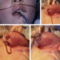
An incision approximately 4 cm in length is marked ( Fig. 1 ) and local anesthetic is injected 2 cm above the costal margin. A subperiosteal dissection begins at the inferior boarder of the costal cartilage and progresses posterolateral until the eighth rib is encountered ( Fig. 2 ). The ribs are dissected to the osseocartilaginous junction laterally using Freer and Doyen rib elevators to carefully separate the cartilage from the intact deep perichondrium. Next, the synchondrosis usually found between ribs 6 and 7 is exposed. The connection between these ribs provides a large enough piece of cartilage to form the base of the framework. The previously made template is placed over the synchondrosis to plan the ideal area of the rib cartilage to harvest, which is then done with the superficial perichondrium intact. A malleable retractor is placed before harvest to protect the pleura and cartilage is cut out ( Fig. 3 ). Approximately 8 cm of eighth rib is needed for reconstruction. The eighth rib is also harvested for the creation of the helix ( Fig. 4 ).
It is important to preserve an intact rim of the superior margin of the sixth rib to prevent chest wall deformity. Maintaining even a minimal rim will anchor the rib to the sternum, preventing chest flair and distortion, something that can become more apparent as the patient grows.
The wound bed is flooded with saline and a Valsalva maneuver is performed to assess for pneumothorax. Bubbles indicate the location of pleural violations. To repair any pleural tears, a soft, red rubber catheter is placed through the pleural rent and a 4-0 chromic purse-string suture is placed around the edges. Suction is applied to the catheter as it is withdrawn and the suture tied. Additional cartilage is banked either under the chest incision or in the scalp for future framework projection in a later stage. Next, the muscle, subcutaneous dermis, and skin of the chest incision are closed in a layered fashion with a suction drain placed over the muscle closure.
The cartilage framework is addressed next. Gentle tissue handling is essential during this stage. The cartilage is living tissue that must be kept moist. Perichondrium should be left intact when possible. Power tools are not used so as to avoid thermal damage to tissue. Use of foreign materials, such as permanent suture, is limited to prevent extrusion.
The framework base is fabricated by carving the sixth and seventh ribs. Sharp blades are used during all stages of carving. After constructing the base shape of the ear, the eighth rib is thinned longitudinally, allowing it to be wrapped around this base and secured judiciously by clear nylon sutures ( Fig. 5 , [CR] ). Exaggerating helical height in this step is important to give the appearance of additional auricular projection.