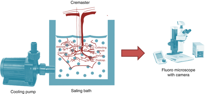
Fig. 18.1
Sketch of isolated cremaster muscle flap with marked arterial and venule branches
The rat is placed in the tissue bath and the muscle spread on coverglass and fixed with sutures. It is transilluminated by fiberoptic lamp and covered by plastic film to keep appropriate moisture. Tissue bath is filled with 0.9 % saline solution and temperature maintained around 36 °C in normothermia or 4 °C in hypothermia. The cooling pump is used to keep the low equal temperature. Fluorescence microscope with color camera is used to observe microcirculation. The system scheme is presented in Fig. 18.2. Following parameters can be measured: arteriolar diameter, red blood cell velocity, leukocyte count in postcapillary venules, capillary density.


Fig. 18.2
Scheme of cooling and monitoring system
Exemplary Experiment
Sixty male Sprague-Dawley rats weighing 130–170 g were randomly assigned to six experimental groups of ten animals each. Anesthesia was induced with intraperitoneal pentobarbital (60–70 mg/kg). The rats were placed on a heating pad, and body temperature (measured by rectal probe) was maintained between 35 and 37 °C. Carotid arterial pressure was monitored throughout the surgical procedure.
The animals were divided into several experimental groups
Group 1: Normothermic Control, +36 °C (ten animals).
Group 2: Hypothermic Control, +4 °C (ten animals).
Groups 3 and 4: Ischemia-reperfusion in normothermia, +36 °C (ten animals). Following flap isolation, vascular clamps were applied to the iliac and femoral artery and vein just above and below the origin of the cremaster pedicle. The flaps were subjected to 4 h (group 3) or 6 h (group 4) of normothermic ischemia. Following clamp release, the microcirculation was observed for an additional 2 h during reperfusion. Measurements were taken at 1-h intervals.
Groups 5 and 6: Ischemia-reperfusion in hypothermia, +4 °C (ten animals). The isolated cremaster muscle flaps were prepared on the hypothermic tissue bath (+4 °C). The flaps were then subjected to 4 h (group 5) and 6 h (group 6) of ischemia followed by 2 h of tissue reperfusion. One of the measured parameters is shown in Table 18.1 [5].
Table 18.1
Arteriolar diameter changes under normal and hypothermic conditions over 4 hours observation period
1 h
2 h
3 h
4 h
Normo
Hypo
Normo
Hypo
Normo
Hypo
Normo
Hypo
Al
130 ± 17
133 ± 33
127 ± 19
110 ± 30
130 ± 15
102 ± 23
136 ± 17
108 ± 24
A2–1
83 ± 20
77 ± 39
83 ± 19
77 ± 29
88 ± 18
69 ± 25
86 ± 21
Stay updated, free articles. Join our Telegram channel

Full access? Get Clinical Tree

 Get Clinical Tree app for offline access


