, Shimin Chang2, Jian Lin3 and Dajiang Song1
(1)
Department of Orthopedic Surgery, Changzheng Hospital Second Military Medical University, Shanghai, China
(2)
Department of Orthopedic Surgery, Yangpu Hospital Tongji University School of Medicine, Shanghai, China
(3)
Department of Microsurgery, Xinhu Hospital Shanghai Jiao Tong University, Shanghai, China
The medial sural perforator flap was described in a very similar fashion in cadaver dissections by Taylor and Daniel [1] as a potential free flap donor site as early as 1975. Baek [2] was the first to report its clinical applications along with anatomical observations regarding medial and lateral femoral free flaps. The true medial sural perforator free flap was first introduced by Cavadas et al. [3].
26.1 Vascular Anatomy
The medial sural artery arises from the popliteal artery and, after running 2–5 cm, enters the deep surface of the medial gastrocnemius muscle. Within the muscle, the medial sural artery may be either a dominant vascular pedicle or may divide into two branches that run longitudinally between the muscle fiber bundles and give off musculocutaneous perforators to the overlying skin [4, 5].
The medial sural artery runs along an imaginary line connecting the midpoint of the popliteal crease and the midpoint of the medial malleolus.
At least one large musculocutaneous perforator exits through the medial head of the gastrocnemius muscle to allow the creation of a true perforator flap using the overlying calf skin territory. The majority of these perforators are clustered in the distal half of the muscle and emanate near the raphe separating the two heads of the gastrocnemius [6, 7].
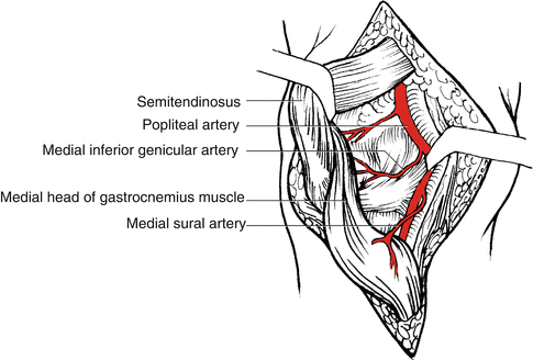

Fig. 26.1
Vascular anatomy of the medial sural artery
26.2 Illustrative Case
A 37-year-old woman suffered a crushing injury to the medial side of her right lower leg, with overlying soft tissue loss measured 6.5 × 5.0 cm (Fig. 26.2).
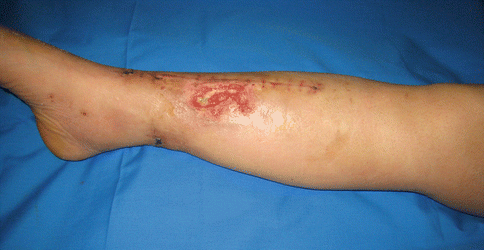

Fig. 26.2
Preoperative view
Flap Design
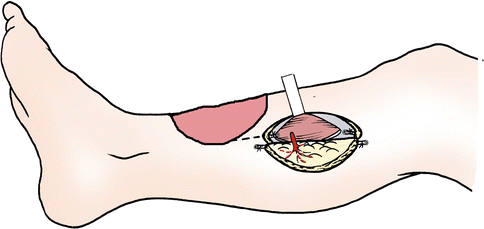
The design of the desired flap is centered around the most distal perforator found to ensure the longest possible pedicle (Fig. 26.3). The main perforators of the medial sural artery are located on a line drawn from the midpoint of the popliteal crease to the midpoint of the medial malleolus. Using an audible Doppler probe to locate a distal perforator over the medial calf, a template of the defect was then centered about this point to create a 6.5 × 4.2 cm flap (Fig. 26.4).
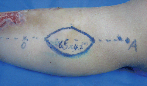
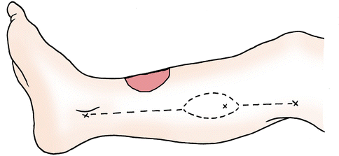

Fig. 26.3
Flap design

Fig. 26.4
Schematic drawing of flap design
Flap Elevation
The lateral and/or distal border of the flap is first raised to confirm the location and size of the perforators (Fig. 26.5). Next, an intramuscular dissection of the identified perforator through the medial gastrocnemius muscle proceeds back to the medial sural vessels to obtain the desired pedicle length. The remaining boundaries of the flap are incised through the deep fascia (Fig. 26.6).
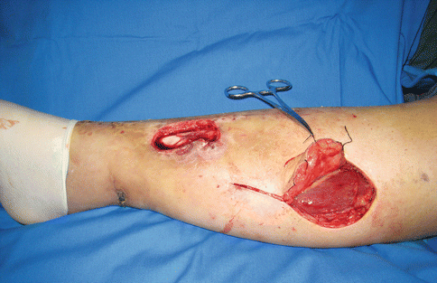

Fig. 26.5
Perforator vessel visualization

