Management of Mandibular Coronoid Fractures
Raja A. Naddaf
PATHOGENESIS
ANATOMY
The zygomatic arch protects the coronoid process along its lateral aspect. The temporalis muscle attaches to the lateral surface of the coronoid process.
PATIENT HISTORY AND PHYSICAL FINDINGS
Clinical exam demonstrates several symptoms, most commonly a limitation in mouth opening and in lateral movement of the mandible.
IMAGING
Plain radiographs or panoramic radiographs can be helpful, but the standard is computed tomography (CT) scans (FIG 1).
DIFFERENTIAL DIAGNOSIS
Considering anatomical and functional relations, when facing a coronoid process fracture, the surgeon must take into consideration the probability of concomitant zygomatic arch fracture.
NONOPERATIVE MANAGEMENT
Treatment of coronoid process fractures can be divided into three categories:
Nonoperative management
Conservative treatment
Open reduction and internal fixation (ORIF)
Nonoperative management is advocated in cases of minimally displaced coronoid process fractures, absence of mouth opening limitation or trismus, and usually when the fracture is solitary.
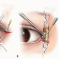
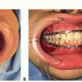
SURGICAL MANAGEMENT
In cases of concomitant zygomatic arch fractures, one should consider closed reduction of the arch fracture only.Related posts:

Stay updated, free articles. Join our Telegram channel

Full access? Get Clinical Tree