Fig. 7.1
Lymphoepithelioma-like carcinoma of the skin. An indurated lesion with a keratotic center on the face
Pathology
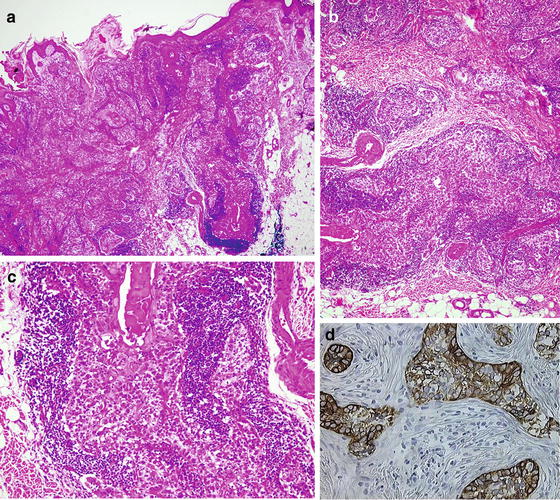
Histologically, LELCS is composed of islands of enlarged atypical polygonal epithelial cells with scant amphophilic to eosinophilic cytoplasm, large vesicular nuclei, and prominent nucleoli surrounded and permeated by a dense lymphoplasmacytic infiltrate (Fig. 7.2a–c). Tumor cells are arranged in round to oval nests, isolated or anastomosing islands, trabeculae, or narrow cords. Mitotic figures ranged from 1 to 8 per high-power field. No connection to the overlying epidermis is present. LELCS exhibits immunoreactivity with high-molecular-weight cytokeratins and epithelial membrane antigen (EMA), indicating the epithelial origin. Neoplastic cells are strongly positive for CK5/6 (Fig. 7.2d), CK14, and CAM5.2 but are negative for CK19 and CK20. The surrounding lymphoid infiltrate exhibits a positive reaction with the T-cell and B-cell markers CD3 and CD20. All tumors show strong p63 protein reactivity. LELCS stains negative to EBV, in contrast to the EBV-positive staining of nasopharyngeal carcinoma. The histogenesis is unclear, although an adnexal origin is favored, with possible follicular, glandular, or sebaceous differentiation. Like undifferentiated nasopharyngeal carcinoma, LELCS could be considered to be a subtype of nonkeratinizing squamous cell carcinoma variant with an epidermal origin.


Fig. 7.2


Lymphoepithelioma-like carcinoma of the skin. (a) A dermal proliferation made by squamous nests in association with clusters of lymphocytes. (b) Squamous nests surrounded by a lymphoplasmacytic infiltrate. (c) Islands of enlarged atypical polygonal epithelial cells with eosinophilic cytoplasm surrounded and permeated by a dense lymphoplasmacytic infiltrate. (d) The epithelial islands are positive for high-molecular-weight cytokeratins
Stay updated, free articles. Join our Telegram channel

Full access? Get Clinical Tree


